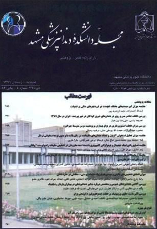
فهرست مطالب

مجله دانشکده دندانپزشکی مشهد
سال چهل و یکم شماره 1 (پیاپی 100، بهار 1396)
- تاریخ انتشار: 1396/02/12
- تعداد عناوین: 10
-
- مقاله پژوهشی
-
صفحه 1مقدمهدر ارتباط با تاثیر سفیدکردن دندان روی خصوصیات مکانیکی مینا نتایج متناقضی وجود دارد. هدف از این تحقیق ارزیابی و مقایسه اثر سفیدکردن در خانه و سفیدکردن در مطب با کمک لیزر روی ریز سختی مینا بود.مواد و روش هادر این مطالعه آزمایشگاهی، 18 دندان انسیزور گاوی سالم انتخاب و به دو گروه تقسیم شدند. گروه اول تحت درمان سفیدکردن در خانه با استفاده از کاربامید پراکساید 15 درصد قرار گرفت (8 ساعت در روز طی 15 روز). در گروه دوم، سفیدکردن در مطب با استفاده از هیدروژن پراکساید 40 درصد انجام گرفت. از لیزر دیود GaAlAs با طول موج nm 810، توان 2 وات و تابش پیوسته در حالت غیرتماسی جهت تسریع سفیدکردن استفاده شد. این روند 3 بار در طی 15 روز انجام گرفت. نمونه ها در طول مطالعه در بزاق مصنوعی Fusayama Meyer نگهداری شدند. سختی نمونه ها به روش Vickers قبل و بعد از درمان های سفیدکردن در هر دو گروه اندازه گیری شد. داده ها با استفاده از آزمون های تی زوجی و تی مستقل ارزیابی و سطح معنی داری 05/0 در نظر گرفته شد.یافته هامیزان سختی مینا بعد از هر دو روش سفیدکردن در خانه و سفیدکردن در مطب به کمک لیزر کاهش یافت، ولی این کاهش معنی دار نبود (05/0نتیجه گیریتحت شرایط این مطالعه آزمایشگاهی، هیچ یک از روش سفیدکردن در خانه و سفیدکردن در مطب با کمک لیزر تاثیر قابل توجهی روی محتوای معدنی مینای سالم نداشتند. بنابراین، هر دو روش را می توان به عنوان روش های محافظه کارانه برای درمان بد رنگی دندان ها توصیه کرد.کلیدواژگان: سفیدکردن دندان، لیزر، ریز سختی، سفیدکردن در خانه، سفیدکردن در مطب
-
صفحه 11مقدمهبا توجه به استقبال عمومی پژوهشگران برای انجام تحقیقات در سال های اخیر، نقد و حذف خطاها در مطالعات یکی از ضرورت ها می باشد.باتوجه به شمار بسیار اندک مطالعات نقد در حیطه دندان پزشکی و به خصوص مطالعات کارآزمایی بالینی دارای گروه کنترل تصادفی شده،مطالعه حاضربه گردآوری این مطالعات ونق دآنها پرداخته است.مواد و روش هامقالات کارآزمایی بالینی دارای گروه کنترل تصادفی منتشرشده در مجله دانشکده دندانپزشکی دانشگاه علوم پزشکی مشهد در سال های 94-1382 گردآوری و بر اساس چک لیست CONSORT امتیازدهیشدند.داده ها با استفاده از نرم افزار SPSS با ویرایش 20 مورد تجزیه و تحلیل قرار گرفتند. شاخص های مرکزی و پراکندگی امتیاز کسب شده محاسبه گردید، همچنین مقالات براساس امتیاز کسب شده به چهار سطح ضعیف، متوسط، خوب و عالی تقسیم بندی شدند.یافته هااز مجموع 514 مقاله، 64 مقاله کارآزمایی بالینی و از این تعداد 40 مقاله کارآزمایی بالینی دارای گروه کنترل تصادفی شده بودند. بیشترین تعداد مقالات کارآزمایی بالینی چاپ شده، 7 مقاله در سال های 1382 و 1388 بود و کمترین تعداد، مربوط به سال های 1383، 1384، 1391 و 1392 بود که تنها یک مقاله از این نوع به چاپ رسانده بودند. میانگین و انحراف معیار درصد امتیازات کسب شده در مجموع مقالات 61/13±79/57 بود. بهترین میانگین امتیاز کل مربوط به سال 1393 بوده است.نتیجه گیریبا توجه به اهمیت و نقش مطالعات کارآزمایی بالینی دارای گروه کنترل تصادفی شده، رفعنواقصذکر شدهوافزایشکیفیتانجام این مطالعات و در نتیجه مقالات حاصله از آن، درکشورضروریبهنظر می رسد.کلیدواژگان: ارزیابی نقادانه، کارآزمایی بالینی، تصادفی سازی
-
صفحه 21مقدمهانجام درمان ارتودنسی می تواند عامل مهمی برای ایجاد پوسیدگی های اولیه باشد. با توجه به خواص ضدپوسیدگی چای سبز، مطالعه حاضر با هدف بررسی توانایی وارنیش آن در پیش گیری از پوسیدگی های ناشی از ارتودنسی ثابت طراحی شد.مواد و روش هادر این مطالعه تجربی، تعداد 45 دندان پره مولر سالم انتخاب و پس از قرار دادن براکت بر روی آن ها به طور تصادفی، به 3 گروه مساوی (15N=) تقسیم شدند. در گروه اول (گروه کنترل)، دندان ها هیچ درمانی دریافت نکردند. در گروه دوم، وارنیش چای سبز در یک دوره 21 روزه، 48 ساعت یکبار در اطراف براکت ها اعمال شد. در گروه سوم، وارنیش چای سبز در یک دوره 21 روزه، 24 ساعت یکبار در اطراف براکت ها اعمال شد. پس از این دوره 21 روزه، مقاطع 40 میکرونی، سرویکالی تر از محل قرارگیری براکت ها تهیه و جهت بررسی پوسیدگی، میانگین عمق ضایعات بر حسب میکرومتر در 3 نقطه به فاصله 500 میکرونی از هم با استفاده از نور پلاریزه، اندازه گیری شد. داده ها توسط نرم افزار SPSS آزمون آماری ناپارامتریک کروسکال والیس و من-ویتنی مورد تجزیه و تحلیل قرار گرفت. سطح معنی دار 05/0 در نظر گرفته شد.یافته هامیانگین و انحراف معیار عمق پوسیدگی گروه اول تا سوم به ترتیب 72/290±03/452، 92/92±91/44 و صفر میکرون بود. گروه اول و سوم به ترتیب بیشترین و کمترین میزان عمق پوسیدگی را دارا بودند. میانگین عمق پوسیدگی در گروه های دوم و سوم تفاوت معنی داری نسبت به هم نداشتند (878/0P=)؛ ولی گروه های اول و دوم و همچنین گروه های اول و سوم اختلاف معنی داری با یکدیگر داشتند (001/0>P).نتیجه گیرینتایج این مطالعه نشان داد که کاربرد وارنیش چای سبز در گروه های مورد مطالعه موجب کاهش معنی دار پوسیدگی دندان در اطراف براکت های ارتودنسی می گردد.کلیدواژگان: چای سبز، وارنیش، ارتودنسی ثابت، لکه سفید
-
صفحه 31مقدمههدف از انجام این مطالعه بررسی شیوع هیپومینرالیزاسیون مولر انسیزور (MIH) در کودکان 13-6 ساله شهر رشت و عوامل پیش بینی کننده آن بود.مواد و روش هاشرکت کنندگان در این مطالعه، 1043 کودک از دبستان های دولتی و غیردولتی رشت بودند. فراوانی MIH براساس معاینه بالینی تعیین گردید و سپس 235 کودک مبتلا به MIH و گروه شاهد آنان جهت تعیین عوامل احتمالی ایجادکننده مورد ارزیابی قرار گرفتند. برای جمع آوری اطلاعات از مصاحبه و پرسشنامه شامل سوالاتی درباره مشکلات اواخر دوران بارداری، هنگام تولد و بیماری های سه سال اول زندگی، استفاده شد. برای بررسی رابطه بین عوامل مختلف از آزمون های کای دو، آزمون دقیق فیشر، من- ویتنی و آزمون تی، استفاده شد (05/0α=).یافته هاشیوع MIH 9/19 درصد و درگیری مولر به تنهایی 46/17 درصد بود. هرکودک به طور متوسط دارای 18/2 دندان دارای ضایعه، شامل 9/1 مولر و 28/0 ثنایا بود. تفاوت معنی داری از نظر شیوع و شدت ضایعات در دو جنس دیده نشد (54/0P=و 36/0=X2)، همچنین بین دانش آموزان مدارس خصوصی و دولتی تفاوت معنی داری از نظر شیوع مشاهده نشد (47/0=X2 و 58/0P=). مولر های پایین بیشتر از بالا دچار نواقص مینایی بودند (6/19= X2و001/0P<). بیشترین فراوانی ضایعات مربوط به لکه های اوپک بود. هیچیک از عوامل مورد بررسی با MIH رابطه معنی داری نداشتند.نتیجه گیریبراساس یافته های این مطالعه، شیوعMIH در شهر رشت از میزان بالایی برخوردار بود ولی بیشتر ضایعات از نوع خفیف و میانگین تعداد دندان های مبتلا در هر فرد، حدود دو دندان بود. بررسی ارتباط دقیق تر با علل اتیولوژیک از جمله عوامل ژنتیک توصیه می شوند.کلیدواژگان: هیپومینرالیزاسیون، مولر، انسیزور، شیوع، اتیولوژی
-
صفحه 41مقدمهتروماهای دندانی یکی از شایع ترین علل مراجعه به مراکز دندانپزشکی می باشد. پیش آگهی تروماهای دندانی، وابسته به اقدامات اساسی بلافاصله بعد از تروما می باشد.هدف از این پژوهش بررسی میزان آگاهی والدین کودکان 12-8 ساله اصفهان در رابطه با ترومای دندان های بیرون افتاده در طی سال 95-94 بود.مواد و روش هادر این مطالعه توصیفی- تحلیلی، بعد از بومی سازی کردن روایی و پایایی آن، تعداد 500 نفر از والدین کودکان 12-8 سال به صورت نمونه گیری دو مرحله(خوشه ای-سهمیه ای) انتخاب شده و به سوالات پرسشنامه جواب دادند. از آزمون های آماری ANOVA و
t-test برای آنالیز داده ها استفاده شد.(05/0=α)یافته هامیانگین مجموع آگاهی والدین برابر با 01/2±25/5 محاسبه شد. بین آگاهی والدین و سطح تحصیلات آنها ارتباط مستقیم و معنی دار ضعیفی وجود داشت (001/0 P< و 165/0=r) ولی بین آگاهی آنان و سن ، جنس و تعداد فرزندان ارتباطی مشاهده نشد. سطح آگاهی والدین در مورد دندان بیرون افتاده در اثر تروما ناکافی ارزیابی شد. 6/44 درصد از والدین اظهار داشتند که دندان دایمی بیرون افتاده را باید جایگزین کرد. 4/10 درصد نفر از آن ها حداکثر زمان جایگذاری دندان بیرون افتاده را 30-20 دقیقه دانستند و همچنین 2/18 درصد شیر و بزاق را به عنوان بهترین محیط جایگذاری دندان انتخاب کردند. اکثریت والدین تلویزیون را منبع کسب اطلاعات خود دانستند.نتیجه گیریوالدین شرکت کننده سطح آگاهی پایینی از تروماهای دندانی منجر به بیرون افتادن دندان از ساکت داشتند. پیشنهاد می گردد که برای افزایش سطح آگاهی والدین برنامه های آموزشی در این ارتباط برگزار گردد.کلیدواژگان: آگاهی، بیرون افتادن دندان از ساکت، کودک، والدین -
صفحه 51مقدمهپرسلن های فلدسپاتیک و سیستم های زیر کونیایی پرمصرف ترین سرامیک های دندانی می باشند. سایش دندان هادر برابر این سرامیک های پرمصرف یک نگرانی عمده می باشد. خشونت سطحی اولیه سرامیک ها می تواند میزان سایش مینای دندان مقابل را تحت تاثیر قرار دهد. هدف از این مطالعه ارزیابی رابطه بین میزان سایش و خشونت سطحی سرامیک های دندانی می باشد.مواد و روش هادر این مطالعه آزمایشگاهی، نمونه های زیرکونیای پالیش شده، پرسلن فلدسپاتیک پالیش شده و پرسلن فلدسپاتیک پالیش و گلیز شده آماده شد (11n=). پرمولر طبیعی انسان نیز به عنوان آنتاگونیست تهیه گردید. سپس از نمونه های دندانی مانت شده توسط استریومیکروسکوپ در موقعیت ثابت عکس گرفته شد و فاصله نوک کاسپ ها تا نقطه مرجع که توسط دیسک روی نمونه دندانی حک شده بود، اندازه گیری شد. خشونت سطحی کلیه نمونه ها قبل از انجام آزمایش توسط دستگاه پروفیلومتر اندازه گیری شد. سپس نمونه های سرامیکی در مقابل نمونه های دندانی مورد آزمایش قرار گرفتند. 11دندان هم به عنوان گروه شاهد در برابر 11 دندان دیگر قرار گرفتند و 120000 سیکل جوشی (معادل 6 ماه جویدن) اعمال گردید. مجددا از نمونه ها عکس گرفته شد. اختلاف این دو مقدار یادداشت گردید. با استفاده از نرم افزار SPSS با ویرایش 20 و آزمون کروسکال والیس و نمودار پراکنش (Scatter) در سطح معنی داری 5 درصد تجزیه و تحلیل آماری انجام شد.یافته هامیانگین و انحراف معیار خشونت سطحی گروه پرسلن فلدسپاتیک پالیش شده 083/0±48/1 میکرومتر و گروه های پرسلن فلدسپاتیک پالیش و گلیز شده، زیرکونیای پالیش شده و گروه کنترل (شاهد) به ترتیب 126/0±20/1، 086/0±535/0 و 006/0±039/0 میکرومتر بود. بین خشونت سطحی گروه های مورد مطالعه تفاوت آماری معنی داری وجود داشت (001/0P<). در بین سرامیک ها بیشترین میزان سایش و خشونت سطحی مربوط به گروه پرسلن فلدسپاتیک پالیش شده و کمترین میزان سایش و خشونت سطحی مربوط به گروه زیرکونیای پالیش شده بود.نتیجه گیریبا افزایش خشونت سطحی سرامیک ها میزان سایندگی آنها نیز افزایش می یابد.کلیدواژگان: سایش مینای دندان، خشونت سطحی، سرامیک دندانی
-
صفحه 61مقدمهتکنیک ساندویچ یکی از روش های متداول در دندانپزشکی است که در آن گلاس آینومرهای تغییر یافته با رزین با توجه به مزیت هایی چون آزاد سازی فلوراید و چسبندگی به ساختمان دندان، همراه با ترمیم های کامپوزیت استفاده می شوند. هدف از این مطالعه بررسی میزان استحکام باند برشی بین گلاس آینومر نوری و کامپوزیت با استفاده از عوامل اتصال دهنده دارای حلال های متفاوت می باشد.مواد و روش هادر این مطالعه تجربی آزمایشگاهی، 80 نمونه گلاس آینومر نوری GC FUJI 2LC آماده سازی شد. در گروه اول بدون استفاده از عامل باندینگ و در سه گروه دیگر با استفاده از عوامل باندینگ Single bond، One step plus و TG bond رزین کامپوزیت به گلاس آینومر متصل شد. پس از یک هفته نگهداری نمونه ها داخل آب مقطر در دستگاه انکوباتور با دمای 5/37 درجه سانتی گراد، توسط دستگاه تست یونیورسال، استحکام باند برشی بر حسب مگاپاسکال اندازه گیری شد. آنالیز واریانس یک طرفه و آزمون LSD جهت تحلیل داده هاو مقایسه بین گروه ها استفاده شد.یافته هابیشترین مقدار استحکام باند مربوط به نمونه های باند شده با عامل باندینگ TG bond (99/12 مگاپاسکال) و کمترین مقدار مربوط به گروه کنترل (3/5 مگاپاسکال) بود. میانگین استحکام باند برشی در چهار گروه تفاوت آماری معنی داری داشت. (001/P=)نتیجه گیریطبق نتایج این مطالعه بیشترین استحکام باند برشی بین گلاس آینومر تقویت شده با رزین و کامپوزیت مربوط به عامل باندینگ TG bond بود که می تواند به دلیل وجود آب در ترکیب باندینگ و اثر آن به عنوان مرطوب کننده گلاس آینومر و به دنبال آن چسبندگی بیشتر کامپوزیت باشد.کلیدواژگان: استحکام باند برشی، کامپوزیت رزین، گلاس آینومر، عامل باندینگ
-
صفحه 69مقدمهاضطراب دندانپزشکی به معنای واکنش روان شناختی بیمار نسبت به استرس ناشی از مداخلات دندانپزشکی است. این نوع اضطراب رایج است و سنجش و ارزیابی آن در جریان درمان های روان پزشکی بسیار سودمند می باشد. هدف پژوهش حاضر، بررسی ویژگی های روان سنجی پرسشنامه اضطراب دندانپزشکی بود.مواد و روش هابه این منظور تعداد 300 نفر از افرادی که سابقه مراجعه به دندان پزشک داشتند، از بین دانشجویان دانشگاه آزاد خوی، به روش تصادفی انتخاب شدند. پس از کسب رضایت نامه آگاهانه جهت شرکت در پژوهش، پرسشنامه اضطراب دندانپزشکی توسط این افراد تکمیل شد. داده های به دست آمده با استفاده از نرم افزار SPSS و LISRELتحلیل شدند.یافته هانتایج تحلیل عاملی اکتشافی، نشانگر استخراج یک عامل عمده بود؛ همچنین ساختار اکتشاف شده برای پرسشنامه اضطراب دندانپزشکی در جمعیت ایرانی از طریق تحلیل عاملی تائیدی، تائید شد. همچنین همسانی درونی این پرسشنامه به روش آلفای کرونباخ (94/0=α) و دونیمه کردن (94/0=r) ارزیابی شد که از میزان بالایی برخوردار بود.نتیجه گیریپرسشنامه اضطراب دندانپزشکی از ویژگی های روان سنجی لازم جهت استفاده در جمعیت ایرانی برخوردار است و می تواند توسط متخصصان برای ارزیابی اضطراب دندانپزشکی مورد استفاده قرار گیرد.کلیدواژگان: اضطراب، دندانپزشکی، روان سنجی
- گزارش مورد
-
صفحه 79مقدمهعلاقه به داشتن دندان های سفیدتر و تکنیک سفید کردن دندان باعث افزایش تقاضای عمومی نسبت به سفیدتر کردن دندان شده است. اصلاح رنگ با سفید کردن اغلب کمترین هزینه را دارد. در این ارتباط، منابع نوری مختلف استفاده می شود این منابع نوری باعث افزایش سرعت اثر مواد بلیچینگ در مطب مثل، هیدروژن پرواکساید، می شود. هنگامی که نور لیزر برای این منظور استفاده می شود، به طور معمول سبب ارتقای جذب نور لیزربه ژل شده و انرژی نورانی تبدیل به حرارتی می گردد که سبب تسریع تاثیر ترکیبات بلیچینگ می شود.
گزارش مورد:در این مطالعه، یک خانم 35 ساله که دچار فلوروزیس دندانی بود، با مشکل بدرنگی دندان ها به دانشکده دندانپزشکی مراجعه کرد. به دلیل گزارش حساسیت شدید دندانی در طی بلیچینگ خانگی، از روش بلیچینگ در مطب با منبع نوری لیزر Er:YAG (Fotona Dualis XS، USA) با توان 4/2 وات کمتر از حد آستانه مینای دندان استفاده شد. تغییررنگدندان ها در حد3-2 درجه، از رنگ A3.5 بهرنگA1در3دقیقهو15ثانی هایجاد شد. به دلیل زمان کار کوتاه و نیز استفاده اولیه از نور لیزر به صورت غیرمتمرکز، بیمار هیچ گونه حساسیتی در حین و پس از کار گزارش نکرد.نتیجه گیریبا توجه به اینکه Er:YAG لیزراستانداردی برای بسیاریازموارد دندانپزشکی است، نیاز به خرید دستگاه لیزر جدید راحذف می کند. بنابراینازلیزر Er:YAG همراهبا موادبلیچینگدرمطب در این مورد به عنوان روشی موثروکمتهاجمجهتسفیدکردندندان ها استفاده شد.کلیدواژگان: فلوروزیس، لیزر، بلیچینگ -
صفحه 83مقدمهسندرم Papillon-Lefevre یک بیماری نادر اتوزومال مغلوب است. این بیماری با هایپرکراتوز کف دست و پا و پریودنتیت سریع پیشرونده مشخص می شود و منجر به از دست رفتن پیش از موعد دندان های شیری و دائمی می گردد. تشخیص و درمان به موقع می تواند مانع از درگیری دندان های دایمی و بهبود پریودنتال و ماندگاری بیشتر دندان های شیری گردد و در پیشگیری از تحلیل ریج استخوانی زودهنگام و ایجاد مشکلات پروتزی برای بیمار کمک کننده باشد.
گزارش مورد: در این گزارش، یک پسر چهار ساله مبتلا به پاپیلون لفور با یافته های هایپرکراتوز کف دست و پا، درگیری شدید پریودنتال و لقی دندان های شیری مورد بررسی و درمان قرار گرفت و بیمار به مدت 4 سال پس از درمان تحت نظر قرار داده شد. با ارائه درمان مبتنی بر شواهد وضعیت پریودنتال بیمار بهبود یافته و امکان ساخت و ارائه پروتز متحرک پارسیل به منظور بازسازی زیبایی و فانکشن بیمار فراهم شد. همچنین وضعیت پریودنتال دندان های دایمی بیمار پس از رویش مطلوب بود.نتیجه گیریبه علت وجود بیماری پریودنتال، دندان پزشکان اغلب اولین کسانی هستند که این بیماری را تشخیص می دهند. شناسایی و درمان زودرس بیماران مبتلا به این سندرم حایز اهمیت می باشد، چرا که می تواند مانع از درگیری دندان های دایمی شود.کلیدواژگان: سندرم پاپیلون لفور، پریودنتیت مهاجم، هاپیر کراتوز
-
Page 1IntroductionResearchers have reported conflicting results in terms of the effects of tooth bleaching on mechanical properties of enamel. This study aimed to evaluate the effect of at-home bleaching and laser-assisted in-office bleaching on microhardness of enamel.Materials And MethodsIn this experimental study, 18 healthy bovine incisors were selected and divided into two groups. Home-bleaching with 15% carbamide peroxide was applied for the first group for eight hours a day over 15 days. On the other hand, in-office bleaching was performed on the samples of the second group using 40% hydrogen peroxide and a GaAlAs diode laser (wavelength 810 nm), applied to accelerate tooth bleaching at settings of 2 W as a continuous wave in non-contact mode. This process was performed for three sessions over 15 days. Specimens were stored in Fusayama Meyer artificial saliva during the study. Vickers hardness test was performed to assess the microhardness of the specimens before and after bleaching in both groups. Data analysis was performed using paired and independent t-tests, and P-value of less than 0.05 was considered statistically significant.ResultsWhile hardness of enamel decreased after both at-home bleaching and laser-assisted in-office bleaching, this reduction was not significant (P>0.05). No significant difference was observed between the study groups in terms of enamel hardness before and after the bleaching procedures (P>0.05).ConclusionAccording to the results of this study, none of the bleaching methods had a significant impact on mineral properties of intact enamel. Therefore, both procedures could be recommended as conservative methods for treatment of discolored teeth.Keywords: Tooth bleaching, laser, microhardness, at, home bleaching, in, office bleaching
-
Page 11IntroductionGiven the considerable attention to research in recent years, review and elimination of errors in any research are of great importance. With respect to the scant review articles within the field of dentistry, especially randomized controlled clinical trials, this study aimed to collect and review these articles.Materials and MethodsAll randomized controlled clinical trials published in the Journal of Dental School, Mashhad University of Medical Sciences, Mashhad, Iran during 2003-2015 were collected and scored based on CONSORT checklist. Data analysis was performed in SPSS version 20. Central tendency and dispersion of the obtained scores were calculated. Accordingly, all articles were classified as weak, medium, good and excellent.ResultsAmong 514 collected articles, 64 were clinical trials, 40 of which were randomized controlled clinical trials. Most clinical trials (seven papers) were published during 2003-2009, whereas only one paper was published in 2012, 2005, 2004 and 2013. Mean and standard deviation of percentage of scores of all articles was 57.79±13.61. In addition, the highest mean score was obtained in 2014.ConclusionAccording to the results of this study, given the importance of randomized controlled clinical trials, it seems necessary to overcome the mentioned shortcomings and increase the quality of these studies.Keywords: Critical evaluation, laser, clinical trial, randomization
-
Page 21IntroductionImplementing orthodontic treatment can cause severe caries in teeth in the shape of light spot. The present study planned to evaluate the ability of green tea varnish in the prevention of caries caused by fixed orthodontic.Materials and MethodsIn the present experimental study, a total number of 45 premolar healthy teeth were selected and categorized into 3 equal groups (N=15) after placing brackets on them. Group 1 (control group): after placing brackets, teeth received no treatment. Group 2: green tea varnish was applied around the brackets in 48 hours intervals for a 21 days period of time. Group 3: green tea varnish was applied around the brackets in 24 hours intervals for a 21 days period of time. After this period, the 40 Micron sections were prepared cervical than the location of the brackets in order to study the avarage of caries depth in micrometers using polarized light in three points at a distance of 500 Micron from each other. The measurment data were analyzed by the SPSS 16.0 software using Kruskal-Wallis and Mann-Whitney nonparametric statistical tests. A significance level of 0.05% was considered.ResultsThe average and standard deviation of the caries depth in groups 1, 2, and 3 is 452.03±290.72, 44.91±92.92 and zero respectively. The control group have the most caries depths compared to other groups. Results of groups 1 and 2 had no significant differenc (P=0.878); but, the groups 1 and 2 and also grous 1 and 3 had significant difference with each other (PConclusionThe results of this study showed that the utilization of green tea varnish in 21-day period at 24 and 48 hours intervals reduces tooth caries around the orthodontic brackets.Keywords: Green tea, varnish, fixed orthodotics, white spot
-
Page 31IntroductionMolar-incisor hypomineralization (MIH) is a rapidly destructive type of enamel defects of systemic origin. The aim of the present study was to investigate the prevalence of MIH and the potential predictive factors among the children within the age group of 6-13 years in Rasht, Iran.Materials and MethodsThe prevalence of MIH was determined through clinical examination of 1043 children aged 6-13 years. At In the next stage, 235 subjects with MIH and their matched controls were evaluated for determining the probable etiologic risk factors. The data were collected using questionnaire and interviews entailing some questions about the late pregnancy problems, birth time problems, and the diseases at the first three years of life. The data analysis was performed using the Chi-square, Fishers exact, and Mann-Whitney U tests through the SPSS version 17. The p-vale less than 0.05 was considered statistically significant.ResultsAccording to the results of the study, the prevalence of MIH was 19.9%, and the sole molar involvement was 17.4%. On average, every child had 2.18 hypomineralized teeth involving 1.9 molars and 0.28 incisor. There was no significant difference between the two genders in terms of the prevalence and intensity of the MIH (P=0.54, X2=0.36). Furthermore, no significant difference was observed between the private and public school students regarding the prevalence of this defect (P=0.51, X2=0.47). As the results indicated, the enamel defects were more common in the lower molars, compared to those in the upper ones (PConclusionAs the findings of the present study revealed, the MIH had a high prevalence. However, most of the defects were mild, and the mean of the MIH-affected teeth was two teeth per child. The opacities were the main type of defect. None of the investigated variables was associated with the MIH. Further studies are recommended to investigate the etiologic factors including the genetic background to determine the predisposing factors.Keywords: Molars, incisors hypomineralisation (MIH), Prevalence, etiology
-
Page 41IntroductionDental trauma is one of the most common causes for visit to the dental office. The prognosis of dental trauma depends on the basic measures taken immediately after the trauma. This study aimed to evaluate the knowledge of parents toward this issue.Materials and MethodsThis cross-sectional study was conducted on 500 parents of children aged 8-12 years, selected using a two-stage sampling (cluster-quota). A standardized questionnaire was prepared and filled by the participants, validity and reliability of which were evaluated after localization. Data analysis was performed using ANOVA and t-test (α=0.05).ResultsIn this study, mean parental knowledge was equal to 5.25±2.01. A slight direct and significant difference was observed between parental awareness and educational level of the parents (r=0.165, PConclusionAccording to the results of this study,low level of knowledge toward traumatic avulsed teeth was observed in the participants. It is recommended that awareness of parents be raised through training programs.Keywords: knowledge, tooth avulsion, Child, parent
-
Page 51IntroductionFeldspathic porcelains and zirconia crowns are the most applied dental ceramic materials. Teeth wear against these ceramics has turned into a major concern. Primary surface roughness of these ceramics can have a significant impact on enamel abrasion. This study aimed to detect an association between enamel abrasion and surface roughness of dental ceramics.Materials and MethodsIn this laboratorial study, polished zirconia samples, polished feldspathic porcelains, and polished and glazed feldspathic porcelains were prepared (N=11). In addition, human natural premolar teeth were provided as antagonist. Afterwards, mounted tooth samples were photographed by a stereo microscope in a fixed position and the distance from the cusp tip to the reference point was measured. Surface roughness of all samples was evaluated before the test using a profilometer device. Following that, ceramic samples were tested against dental samples. Moreover, 11 tooth samples (control group) were tested against another 11 tooth samples in chewing simulator device with 120000 masticatory cycles (equal to six months of chewing). Afterwards, tooth samples were photographed again and differences between the measurements were recorded. Data analysis was performed in SPSS version 20 using the kruskal-Wallis test and scatter diagram graphs, and P-value of less than 0.05 was considered statistically significant.ResultsIn this study, mean and standard deviation of surface roughness rate in polished porcelain, polished and glazed porcelain, polished zirconia ceramics and control group were 1.48±0.083, 1.20±0.126, 0.535±0.086 and 0.039±0.006 micrometers, respectively. According to the results, a statistically significant difference was observed between the level of surface roughness in the study groups (PConclusionAccording to the results of this study, higher level of surface roughness of ceramics increased tooth abrasion.Keywords: Tooth abrasion, surface roughness, dental ceramic
-
Page 61IntroductionSandwich technique is one of the most common methods in dentistry, in which resin-modified glass ionomers are used with composite restorations due to special features, such as fluoride release and adhesion to tooth structure. This study aimed to assess shear bond strength between resin-modified glass ionomer and composite using bonding agents with different solvents.Materials and MethodsIn this in vitro study, 80 resin-modified glass ionomer samples (GC FUJI 2LC) were prepared. Composite was adhered to glass ionomer without the use of bonding agent in the first group and with the use of bonding agents in other three groups (single bond, one step plus and TG bond). After preserving the samples in distilled water in incubator at the temperature of 37.5°C for one week, shear bond strength of the subjects was measured using universal testing machine. Data analysis and comparison of the groups were performed using one-way analysis of variance and tukey post hoc.ResultsIn this study, the maximum amount of bond strength was observed in TG bond group (12.99 Mpa), whereas the lowest amount was found in the control group (5.3 Mpa). Results revealed a statistically significant difference between the groups in terms of mean shear bond strength (P=0.001).ConclusionAccording to the results of this study, the maximum shear bond strength between resin-modified glass ionomer and composite resin was related to TG bond. This could be due to the presence of water in the composition of bonding agent, which acted as a moisturizing agent of glass ionomer and led to greater adhesion of composite to glass ionomer.Keywords: Shear bond strength, composite resin, glass ionomer, bonding agent
-
Page 69IntroductionDental anxiety is defined as psychological reaction to stress caused by dental interventions. This type of anxiety is common among patients, assessment of which could be beneficial in psychiatric treatments. This study aimed to evaluate the psychometric properties of dental anxiety inventory (DAI).
Material &MethodsIn total, 300 students from Islamic Azad University of Khoy, Khoy, Iran with a history of dental treatment were selected by random sampling. Participants filled the DAI after obtaining written informed consents. Data analysis was performed in SPSS and LISREL.ResultsThe results of exploratory factor analysis revealed one factor. In addition, the exploration for DAI in Iranian population was confirmed through confirmatory factor analysis. Moreover, internal consistency of this inventory was demonstrated at Cronbachs alpha (α=0.94) and split-half (r=0.95).ConclusionAccording to the results of this study, DAI contains sufficient psychometric properties for Iranian population and could be used by professionals to evaluate dental anxiety.Keywords: Anxiety, dental, psychometry -
Page 79IntroductionPatient awareness of options available in changing the color of natural dentition has created an increase public demand. Bleaching is the least expensive esthetic treatment option. This process can be performed by several energy sources, which accelerate the effect of bleaching materials, such as hydrogen peroxide, in office. Laser particles used for this purpose usually facilitate the absorption of laser light by the gel, leading to the conversion of light energy to thermal energy causing accelerated bleaching material effects.
Case Report: In this paper, a 35-year-old female patient with a history of dental fluorosis referred to dental college with complaints of tooth discoloration. Given the reports on severe tooth sensitivity during home bleaching, office bleaching was carried out using the laser source of Er:YAG laser (Fontona Dualis XS, USA), with a power of 4.2 watts less than the enamel threshold. This process resulted in 2-3 shade change from A3.5 to A1 after three minutes and 15 seconds of treatment. Patient reported no discomfort or sensitivity during and after the procedure, which was due to the short duration of the process and application of primary use of unfocused laser light.ConclusionAccording to the results of this study, application of Er:YAG laser resulted in the removal of tooth discoloration with bleaching gels in the office. Since the Er:YAG laser is an standard device used for many dental procedures, there was no need for purchasing a new laser device. Therefore, Er:YAG laser could be used along with bleaching gels as an effective and minimally invasive method to whiten teeth.Keywords: Fluorosis, laser, bleaching -
Page 83IntroductionPapillon-Lefevre syndrome (PLS) is a rare autosomal recessive disorder. Clinical manifestations of this disease are hyperkeratosis of the palms and soles and rapidly progressive periodontitis, which result in premature loss of deciduous and permanent teeth. Early diagnosis and correct treatment of this discover could prevent the involvement of permanent teeth and promote periodontal condition, which leads to longer maintenance of deciduous teeth and prevention of alveolar ridge breakdown and prosthetic problems.
Case report: Our case was a 4-year-old boy presented with PLS, hyperkeratosis of the palms and soles and rapidly progressive periodontitis, which had led to the loosening of deciduous teeth. The case has been followed for four years. Evidence-based treatment of the patient resulted in stabilized periodontal condition and the possibility of partial removable prosthodontics for aesthetic and functional reasons. In addition, periodontal condition of permanent teeth of the patient was favorable after the eruption.ConclusionDue to periodontal disease, dentists are often the first to diagnose this syndrome. Early diagnosis and treatment of PLS is of paramount importance due to the possibility of preventing the involvement of permanent teeth.Keywords: Papillon, lefèvre syndrome, aggressive periodontitis, hyperkeratosis


