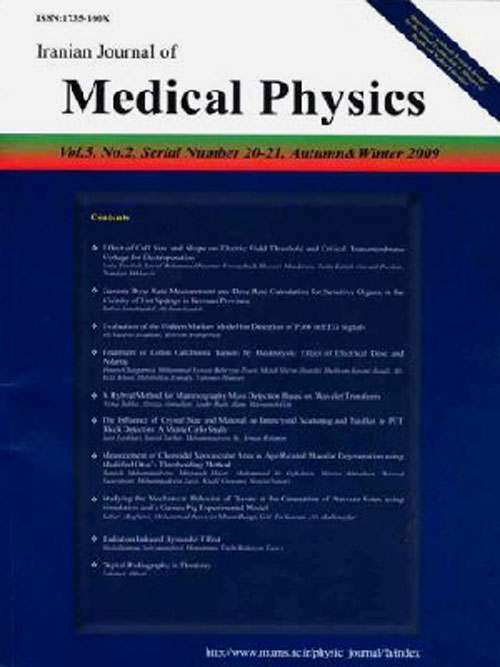
فهرست مطالب

Iranian Journal of Medical Physics
Volume:16 Issue: 2, Mar-Apr 2019
- تاریخ انتشار: 1397/12/10
- تعداد عناوین: 10
-
-
Pages 120-125IntroductionThe present study was conducted to measure the specific activities of 226Ra,232Th and 40K, in some samples of nuts collected from the local markets in Iraq. In addition, this study sought to calculate the annual effective dose of gamma ray to children and adults.Material and MethodsThe quantification of radionuclides was accomplished by gamma spectrometry NaI (Tl) detector.ResultsAccording to the results, the specific activity of 226Ra ranged from 1.39±0.53 to 13.33±1.19 Bq/kg with a mean value of 6.71±1.34 Bq/kg. However, regarding 232Th and 40K, their specific activities had the range values of 0.29±0.09 to 2.43±0.25 and 232.06±8.42 to 376.47±6.26 Bq/kg with the mean values of 1.68±0.50 and 308.57±17.76, respectively. Furthermore, the mean values of the total annual effective radioactive dose in 10-year-old children and adults were 7.43±0.86 and 54.48±6.32 μSv/y, respectively.ConclusionAs the findings indicated, the values obtained for the specific activity of natural radionuclides samples under study were far below the world standard for the ingestion of naturally occurring radionuclide provided by the United Nations Scientific Committee on the Effects of Atomic Radiation (2000) report. The results indicated that the estimated total annual effective radioactive dose in all samples was lower than the value of annual dose limit of 1 mSv/y for public exposure, which is determined by the International Commission on Radiological Protection. Based on the results for each sample, it can be concluded that nut consumption do not expose the Iraqi population to any health risk.Keywords: Iraq, gamma-ray spectrometry nuts, Radioactivity Risk assessment
-
Pages 126-132IntroductionOrgan dose estimation using thermoluminescence dosimeter (TLD) is known to be a standard, although many other methods, such as simulation software, optically stimulated luminescent dosimeters, and photodiodes are still in use. This study aimed at directly measuring mean organ doses to the selected organs in the head/neck, chest, and abdominal regions from four computed tomography (CT) units in Lagos, south-west of Nigeria.Material and MethodsThis study was conducted on locally constructed inhomogeneous phantoms to measure mean organ doses to the head/neck, chest, and abdominopelvic regions from CT units in the Lagos metropolis, Nigeria. Lithium fluoride doped with magnesium and titanium (LiF: Mg, Ti) TLD was used for the measurement. Statistical analysis was performed by IBM SPSS (version 20).ResultsValidation of the designed phantoms was below ± 20% kVp and mAs parameters among the CT units, which was statistically different with regard to the observed dose discrepancies. Generally, a one-way ANOVA showed that there was a statistically significant difference in the investigated mean organ dose (P = 0.043). The comparison of the obtained results from this study with those of other studies revealed that there was no statistically significant difference in the TLDs (P > 0.05). The maximum relative difference in the dose was < 200%.ConclusionThe designed phantoms seemed to be useful for CT dose validation and could be used to validate simulation software in areas where readymade phantoms are not available.Keywords: Thermoluminescent-dosimeter, Computed Tomography Phantom, Organ, organ dose
-
Pages 133-138IntroductionThe aim of the present study was to determine the levels of alpha particles and heavy metals contamination in the meat products consumed in Najaf, Iraq. Moreover, this study was also targeted toward comparing the results with those in the literature and making appropriate conclusion and recommendations.Material and MethodsThis study determined the track of alpha particles and heavy metals pollution in meat samples collected from the open markets in Najaf, Iraq. These meat samples included cow, sheep (lamb), chicken, and fish. The alpha particles contamination was determined using nuclear track detectors (CR-39). The heavy metal concentrations were analyzed using atomic-absorption spectroscopy.ResultsThe highest alpha particles emission rate was 0.0204 mBq cm-2 in Ascary sheep (lamb). On the other hand, the lowest rate of alpha particles (0.00008 mBq cm-2) was associated with Kufa fish. Gadeer sheep and Kufa chicken had the highest and lowest concentrations of cadmium, which were obtained as 0.2600 and 0.0020 ppm, respectively. Regarding the lead concentration, the highest and lowest concentrations were found in Kufa cow (0.8936 ppm) and Kufa chicken (0.0542 ppm), respectively.ConclusionThis study indicated that alpha particle and heavy metal contamination in the meat samples were within permissible limits. Therefore, the consumption of the selected meat products did not pose any significant hazard to the public health in Najaf. Moreover, the findings suggested that there would be no increase in the current rates of not only particle contamination, but also heavy metal pollution, compared to those of international studies.Keywords: Alpha Particles, Atomic Absorption Spectrometry, Environmental Pollution Meat
-
Pages 139-144IntroductionGlobally, intensity-modulated radiation therapy (IMRT) is considered as highly precise and accurate method of radiotherapy planning. This technique amplifies spatial dose distribution conformity by modulating the intensity of radiation beams in each sub-volume. Additionally, it can reduces the dose to surrounding critical organs and deliver the planned dose to targets with the nominal risk of side effects.Material and MethodsIn this study, 13 patients with head and neck cancer were randomly taken for analysis. The IMRT and Rapid Arc plans were generated for each case in the Eclipse treatment planning system, version 11.0. There were seven to nine beams deployed in IMRT plan, while Rapid Arc plans were performed using two arcs with opposite direction of rotation. Portal dosimetry plans were created and analyzed before executing the plan on the patient.ResultsThe mean of V95%(Target’s volume covered by 95% isodose line)was 97.89% and 97.47% for Rapid Arc and IMRT plans, respectively. Moreover, mean standard deviations were found 1.93 and 1.70 in Rapid Arc and IMRT plans, respectively. The mean gamma index was97.55% and 98.43% in Rapid Arc and IMRT, respectively.ConclusionIMRT technique was slightly better in the treatment of head and neck cancer compared to the Rapid Arc method. The only advantage of Rapid Arc was saving the treatment time by two to three times on an average compared with IMRT. It is prudent to use IMRT technique in head and neck cancer treatment.Keywords: Dosimetry, Beam, Cancer
-
Pages 145-151IntroductionThe present study was conducted to implement a simple practical independent quality check of depth dose and isotropy of the Intrabeam™ therapeutic X-ray machine using radiochromic EBT2 film.Material and MethodsTheindependent quality check of 1.5, 3.5, and 5-cm spherical Intrabeam™ applicators was accomplished using particular EBT2 film cutting pieces with internal rounded edges in a water phantom. Prior to this measure, the film was calibrated at three distances from the 5-cm applicator in water to clarify the effects of beam spectrum and dose rate alteration on film response. To this end, three calibration curves were plotted.ResultsThe results of the one-way analysis of variance showed a critical difference between film pieces receiving equal doses at various distances (P<0.05). Therefore, depth dose curves were designed using all three calibration curves. Smaller applicators represented steeper dose fall-off, compared to the larger sizes. In this regard, 14.97%, 17.59%, and 30.92% of the relative mean doses were measured at 1 cm depth of 1.5-cm, 3.5-cm, and 5-cm applicators, respectively. A 10%/1mm gamma index was satisfied for the lateral dose evaluation of corresponding depth relative to Z-direction.ConclusionThe approach implemented in this study could be carried out as a rapid monthly quality check method for the dose distribution evaluation oftheIntrabeam device.Keywords: Analysis of Variance, Calibration, x-rays, water
-
Pages 152-157IntroductionRadioisotopes are naturally the main sources of human exposure to external and internal radiation. Biscuit is a type of food that is widely distributed in all markets, especially in the markets of Iraq. Therefore, the current study aimed to measure the radiation level of some nuclei in biscuit samples and determine the radiation risks that may be caused by this snack.Material and MethodsThis study aimed to evaluate the concentration of alpha radiation activity in 22 different samples of biscuits collected from the markets in Iraq. The analysis of radium activity and radon exhalation rate was performed by employing alpha-sensitive CR-39 plastic track detectors.ResultsThe effective radium values ranged within 23.312-200.44 Bq/kg with a mean value of 58.927 Bq/kg. Radon emission values for the mass unit was within the range of 0.172-1.515 𝐵𝑞/kg.h, with a mean of 0.445 Bq /kg.h, while radon emission values for the surface unit were 3.988-34.3 𝐵𝑞 /𝑚2.h, with a mean of 10.081 Bq /m2.h. The uranium concentrations found in these samples were within the range of 0.02-0.172 ppm with a mean value of 0.05 ppm. Moreover, there was a direct relationship between radium activity and radon exhalation rate. Additionally, the findings showed that uranium correlated positively with radium activity.ConclusionThe results of the present study were within internationally permissible limits. Therefore there is no risk of consumption of biscuits on human health. However, we must use modern techniques and techniques to reduce radiation risk.Keywords: Uranium, radium, Radon, CR-39
-
Pages 158-165IntroductionThis study investigated the effects of extremely low frequency electromagnetic fields (ELF-EMF) (50 Hz, 3 mT) on biochemical parameters of rats’ ovarian tissues and the impact of Allium cepa on the reduction of potential adverse influences of electromagnetic exposure.Material and MethodsIn this study 40 female Wistar rats were divided into four groups, including (1) control group (with 3 cc normal saline), (2) ELF-EMF group (exposed to ELF-EMF, 50 Hz), (3) Allium cepa group (received 3 cc Allium cepa), and (4) ELF-EMF and Allium cepa group (exposed to ELF-EMF and simultaneously received Allium cepa daily for 6 weeks.ResultsThe MDA levels significantly increased in the second group, which were exposed to ELF-EMF and decreased in normal rats that received Allium cepa. Although, SOD, GPx, and CAT activities significantly decreased in ELF-EMF group, the combination treatment with Allium Cepa on exposed rats restored their activities to normal levels. The conduction of transmission electron microscopy study on ELF-EMF group revealed the changes regarding cytoplasmic organelles in the ovarian follicles of exposed rats. Moreover, irregular oocyte with damaged heterochromatic nuclei was observed. In degenerative oocyte, mitochondria lost their cristaeConclusionThe results of the present study suggested that ELF-EMF exposure might cause deleterious effect on ovarian tissues in rats, which may lead to infertility and subfertility. Moreover, using Allium cepa as a nutritional supplement can have beneficial effects in the protection of biological antioxidants and reproductive systems in cases exposed to ELF-EMF.Keywords: Antioxidant Electromagnetic Fields Ovary, Transmission electron microscope
-
Pages 166-170IntroductionThe purpose of this study is to investigate the effects of grid and non-grid techniques in the lateral cervical spine radiography on image quality and entrance surface dose (ESD). Although image quality and radiation doses have been studied by researchers, there is still a dearth of information on image quality and patient dose with different techniques.Material and MethodsThe radiographs of the lateral cervical spine were acquired by positioning the RANDO phantom abutting the erect bucky while using the grid and non-grid techniques. This study benefited from using a 24 cm x 30 cm Fuji standard cassette type imaging plate. A Leeds TOR test tool was utilized for relative comparison of image quality. The ESD of each examination was determined by using the optically stimulated luminescence dosimeter.ResultsThe increased kilovoltage (kVp) resulted in the reduction of ESD whether moving grid, stationary grid, or non-grid techniques were utilized. Significant differences in terms of contrast sensitivity and spatial resolution were indicated when comparing the grid technique to that of the non-grid technique (i.e., χ2=8 and 5, 16 respectively, p<0.05"> ). The results also indicated significant differences in ESD when using the moving grid, stationary grid, and non-grid techniques (i.e., χ2=7.2, 16p<0.05"> ).ConclusionSignificant differences in image quality and ESD were indicated when grid and non-grid techniques were used in the lateral cervical spine radiography. A non-grid with the highest appropriate kVp is recommended as the air gap acts as a grid, resulting in acceptable image quality with reduction in ESD.Keywords: cervical vertebrae, Digital Radiography, Image Quality, Radiation Dosage
-
Pages 171-178IntroductionThis study examined the dosimetric effects based on the rotational setup error to correct patient setup errors occur during volumetric modulated arc radiotherapy (VMAT) for brain tumor patients.Material and MethodsThis study included 1129 cases of cone beam computed tomography (CBCT) images obtained from 46 brain tumor patients, who experienced VMAT and used the 6DoF (degree of freedom) treatment couch. The dosimetric effects regarding the application of the rotational setup error were examined by comparing the treatment plans.ResultsThe mean patient setup errors at the lateral (X-axis), longitudinal (Y-axis), and vertical (Z-axis) directions were 0.1±1.4, 0.0±1.1, and -0.4±1.2 mm, respectively. The pitch, roll, and yaw were -0.29±0.61°,-0.42±0.98°, and -0.53±0.69°, respectively. When an absolute value was taken for the setup error, the mean error was 1.06±0.14 mm at the three translation directions, and the error of rotation was 0.82±0.14°, showing a larger error than that of translation. In terms of the mean dose difference by each region of interest (ROI) before and after correcting for the rotational setup error on the treatment plan, Brain_max was 2.17 Gy, and Brain_mean was 0.28 Gy, whereas the maximum and mean of Brain_stem were -3.58 and -4.43 Gy, respectively. These findings suggested a dose difference according to the correction of the rotational setup error.ConclusionThis study indicated that the dose effect is influenced by the rotational setup error in VMAT of brain tumor patients. Moreover, the 6DoF positional correction could reduce the positional uncertainties and deliver a more accurate dose.Keywords: Volumetric modulated arc radiotherapy, Cone beam computed tomography, Setup error, Dose
-
Pages 179-188IntroductionHeterogeneity correction is an important parameter in dose calculation for cancer patients where it may be cause inaccuracy in dose calculation as a result of different densities of patients. This study studied the impact of dose calculation of breast cancer patients with and without heterogeneity correction.Material and MethodsTwenty breast cancer patients were treated with Three-Dimensional Conformal Radiotherapy(3DCRT). Dose calculations were performed using two modes: Fast Photon mode for homogeneity and Fast Photon Effective Path length for heterogeneity with two photon energies. Monitor Units(MU), Modulation Factor, Dose Volume Histograms(DVH) and quality indices were used to evaluate the effect of heterogeneity correction on dose calculation and investigate the mechanism of this effect in the low and high energies.ResultsHeterogeneity correction compared to without it showed significant reduction in MU and modulation factor at 6MVand 10MV (p<0.05). Dosimetric parameters derived from DVH were significantly lower for Planning Target Volume (PTV) with homogeneity versus heterogeneity (p<0.05) as D95% (95.1%vs93.7%) and V95%(95.3%vs89%) for 6MV while max Dose and D2 increased. Also the dose for organs at risk exhibited an increase with heterogeneity correction. Quality indices were be worst with heterogeneity correction with a significant difference (p <0.05). The differences between the dose with heterogeneity correction and without it in 6MV and 10MV were as follows: ΔD95% (4.4%vs3.4%;P=0.001) and ΔV95%(4.76%vs4.5%;P=0.001).Conclusionnon-use of the heterogeneity correction can be cause to deliver under or overdose dose to the target volume. Tissue heterogeneity correction had an impact on dose calculation for breast cancer patients and this impact was more effective for the low energy.Keywords: Breast Cancer, Conformal Radiotherapy Heterogeneity Correction Photon Energies, Treatment Planning

