فهرست مطالب

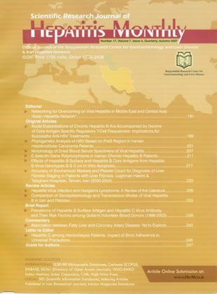
Hepatitis Monthly
Volume:7 Issue: 4, Autumn 2007
- 75 صفحه،
- تاریخ انتشار: 1386/11/16
- تعداد عناوین: 13
-
-
Page 183Background And AimsAcute exacerbations (AEs) of perinatally-acquired chronic hepatitis B (CHB) are accompanied by increased T cell responses to hepatitis B core and e antigens (HBcAg & HBeAg). Naturally-arising forkhead transcription factor Foxp3 (forkhead box p3)-expressing CD4+CD25+ regulatory T (Treg) cells are thought to be important in the control of infectious diseases. This study aimed to investigate whether HBcAg-specific Treg cells play a role in modulating spontaneous AEs and in influencing the outcome of anti-hepatitis B virus (HBV) treatments.MethodsThe SYFPEITHI scoring system was employed to predict epitope peptides on HBcAg overlapping with HBeAg for the construction of peptide-HLA class II tetramers to measure HBcAg-specific Treg cell frequencies (Treg f).ResultsHBcAg-specific Treg f declined significantly in association with increased HBcAg-specific cytotoxic T lymphocyte frequencies during spontaneous AEs without treatment. Vigorous in vitro expansion of CD4+CD25+ Treg cells from CHB patients responding to HBcAg and/or its peptides plus interleukin-2 (IL-2) was consistently detected. Depletion of Treg cells from peripheral blood mononuclear cells enhanced proliferation to HBcAg. In contrast, patients with AEs who received anti-HBV treatments with oral nucleoside analogues or interferon-alpha injection revealed that more post-treatment increase of HBcAg-specific Treg f correlated with a higher sustained remission rate to the therapy.ConclusionsThese data indicate that HBcAg-specific Treg cells from perinatally-acquired CHB patients are proliferative to HBcAg and its peptides and exhibit suppressor activity. They play a crucial role in modulating spontaneous AEs and in successful anti-HBV treatments.
-
Page 201Background And AimsThere are eight genotypes (A-H) of hepatitis B virus (HBV), which show a characteristic worldwide distribution. Genotyping can be accomplished based on a partial sequence of HBV genome such as the PreS or S gene. The aim of this study was to determine the HBV genotypes in Iranian hepatocellular carcinoma (HCC) patients with chronic HBV infection.MethodsSerum sample of 10 HCC patients with chronic HBV infection were subjected to PreS Hemi-Nested PCR. The viral genotype of each sample was determined by bi-directional sequencing of the PreS amplicon and phylogenetic analysis by comparing the nucleotide sequence with 33 reference HBV strains obtained from the GenBank.ResultsPhylogenetic analysis based on PreS region sequences disclosed that all isolated strains belonged to genotype D. Analysis of sequences revealed that all the sequences contained amino acid substitutions. In the PreS2 region of two samples, a point mutation in the start codon was found. There were some deletions with 3, 6 and 8 amino acids in PreS2 region of three samples.ConclusionsDespite the low number of samples, these data revealed that the HBV genotype D is dominant in Iranian HCC patients. Most of the mutations are located at immunodominant epitopes involved in B or/and T cell recognition.
-
Page 207Background And AimsFree and initiated crystallogenesis characteristics of blood serums of 32 healthy people, 14 patients with viral hepatitis B and 12 patients with viral hepatitis C were studied. Specific characteristics of each hepatitis type were singled out.MethodsClassic crystallography and comparative tezigraphy were performed on blood serums. Crystalloscopic fascia was described by identification table and additional criteria. Tezigraphic component was analyzed by basic and additional criteria.ResultsIt was determined that the character of blood serum free crystallization of viral hepatitis is considerably differing from the biological fluid fascia received from healthy people. Significant differences are detected between mounts of control group people and patients with hepatitis B according to the basic tezigraphic coefficient with the initiator potential growth (P<0.01).ConclusionsWe established that it is quite possible to use crystallodiagnostics of the viral hepatitis B and C by the analysis of blood serum fascias.
-
Page 211Background And AimsThe aim of this study is to detect the substitutions Ser128Arg (A128C) and Leu554Phe (T554C) which responsible for E-selectin polymorphisms in patients with chronic hepatitis B and healthy controls. We investigated association of the Ser128Arg, Leu554Phe gene polymorphisms in the E-selectin gene as prototypical inflammatory molecules for susceptibility to chronic hepatitis B.MethodsSixty-three patients with chronic hepatitis B virus infection and 150 healthy subjects were recruited sequentially as they presented to clinic. Genomic DNA was isolated from anti-coagulated peripheral blood Buffy coat using Miller''s salting-out method. The presence of the E-selectin gene polymorphisms was determined by using polymerase chain reaction amplification refractory mutation system (ARMS).ResultsDistribution of E-selectin 128 (A+C-, A+C+, A-C+) genotypes and E-selectin genotype 554 (C+T-, T+C-, C+T+) genotypes were not statistically significant different in chronic hepatitis B patients and controls by Chi-square test (P=0.41 & 0.96, respectively). Also, Two groups had not statistically significant difference in distribution of frequencies of allele 128 A by Chi-square test (P=0.41), 128 C (P=0.15), allele 554 C (P=0.85), and allele 554 T (P=0.76). Carrying allele 128 A (OR=0.587, 95% CI=0.162-2.124), 128 C (OR=1.526, 95% CI=0.849-2.745), 554 C (OR=1.245, 95% CI=0.128-12.089), and allele 554 T (OR=0.880, 95% CI=0.384-2.014) were not risk factors for susceptibility to chronic hepatitis B infection.ConclusionsCarrying E-selectin gene polymorphisms of Ser128Arg and Leu554Phe are not risk factors for susceptibility to chronic hepatitis B.
-
Page 217Background And AimsTo initially explore the underlying pathogenesis of the relationship between genotypes of hepatitis B virus (HBV) and its clinical manifestations.MethodsThe S and C genes of HBV from 60 serum samples, infected by HBV of genotypes B or C were amplified by PCR. The products were recombined with vector pEGFP-C1, which is an internal reference for transfection, to construct the eukaryotic expression recombinant plasmids, followed by cloning and subcloning. Then they were transfected into hepatocarcinoma cell HepG2. The increment rates and apoptosis rates of these transfected cells were determinated by MTT and flow cytometer, respectively.ResultsThe 120 eukaryotic expression recombinant plasmids were all constructed successfully. As an internal reference for transfection, EGFP confirmed that large S protein and C protein of HBV had been expressed in all HepG2 cells. It was found by flow cytometer that the apoptosis rates of HepG2 cells transfected by pEGFP-C1/HBs or pEGFP-C1/HBc from HBV-genotype C samples were all significantly higher than that from HBV-genotype B samples (P=0.009 & P=0.001, respectively).ConclusionsHBV of genotype C can induce more serious cell apoptosis than HBV of genotype B. Difference in apoptosis may be an important reason that HBV of genotype C can induce more severe liver injury than HBV of genotype B.
-
Page 223Background And AimsLiver biopsy is the best known technique for evaluation of liver fibrosis. However, alternative diagnostic methods have been investigated to replace this invasive procedure. Our aim was to assess the diagnostic performance of platelet count and biochemical markers for the staging of liver fibrosis in our patients.MethodsIn a descriptive study, records of all consecutive patients who underwent liver biopsy at Loghman Hakim & Taleghani hospitals between March 2000 and March 2004 were retrospectively studied. The clinical and laboratory data were obtained from patients'' clinical records. Liver histology samples were reviewed by an expert pathologist. The platelet count, serum albumin levels, and the ratio of serum aspartate aminotransferase (AST) levels to platelet count were used as markers for fibrosis. Cutoff points for some of these markers were established using the ROC curve.ResultsOne hundred thirty patients (94 males & 36 females) were studied. The AST/platelet ratio at the cutoff point 0.39 had a positive predictive value (PPV) of 0.70 and a negative predictive value (NPV) of 0.54 to differentiate patients without liver fibrosis from those with moderate fibrosis. This ratio had a PPV = 0.76 and NPV = 0.70 to differentiate patients without liver fibrosis from those with severe fibrosis when used at a cutoff point of 0.25. The platelet count at the cutoff point of 158500 was used for the distinction of mild from moderate fibrosis. The platelet count at the cutoff point of 151000 and serum albumin at the cutoff point of 3.6 were used for distinction of mild from severe fibrosis. No single marker was found to diagnose different stages of fibrosis.ConclusionsThe AST/platelet ratio had an appropriate cutoff point to differentiate patients without liver fibrosis from patients with moderate to severe liver fibrosis. The clinical utility of these tests requires further investigation.
-
Page 229Hepatitis virus infection is an increasing problem, with millions of people all over the world being infected. It is accepted as a significant public health problem with several life altering complications, especially hepatocellular carcinoma. Hepatitis viruses especially for hepatitis C and hepatitis G have been mentioned as a risk factor for development of Hodgkin''s lymphoma. In this work, the author summarized the evidence on the correlation between Hodgkin''s lymphoma and hepatitis virus infection focusing on hepatitis C and hepatitis G. Conclusively, hepatitis C and hepatitis G virus can contribute high risk for development of Hodgkin''s lymphoma.
-
Page 233Hepatitis B virus (HBV) infection is endemic in the Middle East region and is associated with significant morbidity and mortality. Strict strategies are needed for prevention, diagnosis and management of HBV infection. Reviewing literature about seroepidemilogy and modes of infection transmission in Iran and Pakistan performed. Iran is in low endemicity and Pakistan in intermediate endemicity of HBV infection, now. Therapeutic injections, vertical transmission, transfusion, cultural and special traditions like ear, nose piercing, and high risk groups are important risk factors in Pakistan. Prevalence of HBV infection is still significant in children. High risk behaviors, including injection drug use (IDU) and sexual contact are main routes of HBV transmission in Iran. Intensifying vaccination of high risk groups and control on interfamily transmission in both countries is necessary. Effective coverage of HBV vaccination, has more control on therapeutic injections, screening pregnant women for HBV infection, and follow-up of babies of the HBsAg positive mothers in Pakistan is recommended. Regional collaboration of the two countries may overcome the spread of infection by promoting universal vaccination in all provinces of Pakistan, screening of hepatitis B, education, and surveillance in high risk groups of Iran. To implicate effective vaccination by regional and international health units, and addiction control in neighboring countries is necessary.
-
Page 239Background And AimsMillions of lives are saved each year through blood transfusion. Although blood is a life-saving element, it can occasionally cause some severe diseases. This study was performed to assess the prevalence of hepatitis B and C virus infections and their known risk factors among Guilan''s volunteer blood donors from 1998 till 2003.MethodsThe study population consisted of 221,508 blood donors referring to the Blood Transfusion Organization, Guilan, Iran. Enzyme-linked immunosorbent assay (ELISA) was performed for hepatitis B surface antigen (HBsAg) and hepatitis C virus antibody (HCV-Ab) detection. Positive cases were confirmed by neutralization and Recombinant Immunoblot Assay (RIBA), respectively. Known risk factors including histories of surgery, icterus, blood transfusion, endoscopy, unsafe sexual contact, etc. were extracted from available files and evaluated.Results997 individuals were positive for HBsAg and 3,603 individuals for HCV-Ab. After confirmation tests, the prevalence of HBsAg and HCV-Ab was 0.45% and 1.62%, respectively. The most common risk factors were history of surgery followed by icterus in cases or their family.ConclusionsThe prevalence of HBsAg and HCV-Ab is less than that of normal population due to careful screening carried out by staff of Blood Transfusion Organization. Regarding the high frequency of surgery history in positive cases, attending to hospital and operation room hygiene seems to be very important.
-
Guide for AuthorsPage 247


