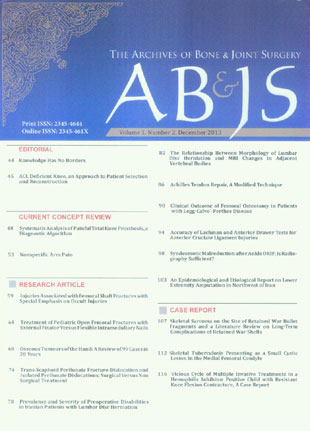
فهرست مطالب

Archives of Bone and Joint Surgery
Volume:1 Issue: 2, Mar 2013
- 124 صفحه،
- تاریخ انتشار: 1392/09/14
- تعداد عناوین: 18
-
-
Page 44
-
Page 48Remaining pain after total knee arthroplasty (TKA) is a common observation in about 20% of postoperative patients; where in about 60% of these knees require early revision surgery within five years. Obvious causes of this pain could be identified simply with clinical examinations and standard radiographs. However, unexplained painful TKA still remains a challenge for the surgeon. The management should include a multidisciplinary approach to the patient`s pain as well as addressing the underlying etiology. There are a number of extrinsic (tendinopathy, hip, ankle, spine, CRPS and so on) and intrinsic (infection, instability, malalignment, wear and so on) causes of painful knee replacement. On average, diagnosis takes more than 12 months and patients become very dissatisfied and some of them even acquire psychological problems. Hence, a systematic diagnostic algorithm might be helpful. This review article aims to act as a guide to the evaluation of patients with painful TKA described in 10 different steps. Furthermore, the preliminary results of a series of 100 consecutive cases will be discussed. Revision surgery was performed only in those cases with clear failure mechanism.Keywords: Diagnostic algorithm, Failure analysis, Pain, Total knee arthroplasty
-
Page 53Nonspecific activity-related arm pain is characterized by an absence of objective physical findings and symptoms that do not correspond with objective pathophysiology. Arm pain without strict diagnosis is often related to activity, work-related activity in particular, and is often seen in patients with physically demanding work. Psychological factors such as catastrophic think- ing, symptoms of depression, and heightened illness concern determine a substantial percentage of the disability associated with puzzling hand and arm pains. Ergonomic modifications can help to control symptoms, but optimal health may require collaborative management incorporating psychosocial and psychological elements of illness.Keywords: Arm, Idiopathic arm pain, Nonspecific arm pain, Shoulder
-
Page 59BackgroundFractures of the femoral shaft are mostly the result of high-energy accidents that also cause multiple trauma injuries, in particular ipsilateral knee and hip injuries.The purpose of this study was to investigate the incidence of injuries associated with femoral shaft fractures and how many of them were undetected.MethodsWe studied 148 patients (150 femoral shaft fractures) with an average age of 52 (range: 18-97). Femoral shaft fractures were treated with antegrade intramedullary nailing in 118 cases (78.7%), and with open reduction and internal fixa- tion in 32 cases (21.3%). Unlocked reamed intramedullary nailing was performed in Winquist type I and type II fractures, while statically locked unreamed intramedullary nailing was carried out in Winquist type III and type IV fractures.ResultsThere were 70 patients with associated injuries (46.4%). The associated injuries went undetected in 18 out of 70 patients (25.5%). Six femoral nonunions (4%) occurred in patients under 70 years of age (high-energy accidents) treated by open reduction and internal fixation.ConclusionInjuries associated with femoral shaft fractures were very frequent (46.4%) in our series, with 25.5% unde-tected. Open reduction and internal fixation was a poor prognostic factor of nonunion in these fractures.Keywords: Associated injuries, Diaphysis, Femur, Fracture, Treatment
-
Page 64BackgroundIn children, inappropriate treatment of open femoral fractures may induce several complications. A few stud- ies have compared the external fixator with flexible intramedullary nails in high-grade open femoral fractures of children. The present study aims at comparing results of these two treatment methods in open femoral fractures.MethodsIn this descriptive analytical study, 27 patients with open femoral fractures, who were treated using either the external fixator (n=14) or TEN nails (n=13) method from 2006-2011, were studied. Some patients were treated with a com- bination method of TEN and pin. The results were evaluated considering infection, union, malunion, and refracture and the patients were followed up for two years.ResultsMean time required for fracture union was 3.89 (range: 2-5.8) and 3.61 (range: 2-5.6) months for the external fixa- tor and TEN groups, respectively. The difference was not statistically significant and there was not any significant difference between the two groups considering infection of the fractured area. Osteomyelitis was not observed in any group. There was an infection surrounding the external fixator pin in 4 cases (28.5%) and so this required changing the location of the pin. In the TEN group, one case (7.6%) of painful bursitis was observed at the entry point of TEN and so the pin was removed earlier than usual. There were two cases (14.2%) of femoral refracture in the external fixator group. Malunion requiring correction was not observed in any of the groups. There were no complications observed in five patients treated with a combined method of pin and flexible intramedullary nails.ConclusionBoth external fixator and intramedullary nail methods are effective ways in treating high grade open femoral fractures in children and final treatment results are similar. Combining pins and flexible intramedullary nails is effective in developing more stability and is not associated with more complications.Keywords: Children, Diaphyseal open fractures, External fixator, Flexible intramedullary nail, Fracture fixation
-
Page 68BackgroundTumors involving the hand skeleton are rare. However, a basic knowledge of hand tumors is necessary for every clinician. This is due to the importance of distinguishing typical benign tumors from life or limb threatening malignant ones.MethodsThis study is a review of 99 cases of osseous hand tumors presented to the department of orthopedic surgery, Imam Khomeini Hospital in Tehran, Iran, from December 1990 to February 2011.ResultsNinety-one cases were benign osseous tumors of the hand and eight tumors were malignant which four of them were considered as primary and four considered as metastatic type. The most common benign tumors were enchondroma and osteoid osteoma. Other benign tumors were epidermoid bone cyst, giant cell tumor of the bone, aneurysmal bone cyst, osteoblastoma, and osteochondroma. Primary malignant tumors were extremely rare and we have reported two chondrosar- comas, one osteosarcoma and one Ewing’s sarcoma involving the hand skeleton.ConclusionThis study indicates that the history, physical examination, laboratory and radiographic data as well as clini- cians’ knowledge of specific hand tumors are required for the best management strategy. New techniques could lead to earlier diagnosis, prevent complications and indentify the most effective type of treatment.Keywords: Benign bone tumors, Hand tumors, Malignant bone tumors, Surgical management
-
Page 74BackgroundTrans-scaphoid perilunate fracture-dislocation and perilunate dislocations are among uncommon injuries, most commonly seen in young patients due to high energy trauma. The treatment can be achieved either surgically by open reduction and internal fixation or closed reduction and casting.MethodsTo compare surgical versus non-operative results of treatment of trans-scaphoid perilunate fracture-dislocation and isolated perilunate dislocation, we collected the data of 34 patients who were treated at least 5 years before our study, twenty of whom were treated surgically and fourteen were treated nonsurgical. We compared clinical and radiological findings in two groups. Functional outcome was assessed by Mayo wrist score for each patient.ResultsThe surgically treated patients had much higher Mayo wrist scores, 85 and 87.78 for perilunate dislocation and trans-scaphoid perilunate fracture-dislocation respectively, while 71 and 71.11 in non-surgically treated group respectively. Wrist range of motion was also more favorable in operative group (55 flexion - 54.28 extension for trans-scaphoid perilunate fracture-dislocation and 50 flexion, 51.66 extension for perilunate dislocations)than non-operative group(48.5 flexion, 48.1 extension for trans-scaphoid fracture-dislocations and 48.1 flexion, 50 extension for perilunate dislocation). The radiographic changes showed arthritic changes but those changes did not significantly interfered with functional outcome and wrist scores.ConclusionRegarding our better clinical results after early open reduction and internal fixation for these injuries, we can suggest the operativetreatment of these complicated hand injuries.Keywords: Hand surgery, Non, operative treatment, Open reduction, internal fixation, Perilunate dislocations, Trans, scaphoid fracture, dislocation
-
Prevalence and Severity of Preoperative Disabilities in Iranian Patients with Lumbar Disc HerniationPage 78BackgroundLiterature recommends that refractory cases with lumbar disc herniation and appropriate indications are better to be treated surgically, but do all the patients throughout the world consent to the surgery with a same disability and pain threshold? We aim to elucidate the prevalence and severity of disabilities and pain in Iranian patients with lumbar disc herniation who have consented to the surgery.MethodsIn this case series study, we clinically evaluated 194 (81 female and 113 male) admitted patients with primary, simple, and stable L4-L5 or L5-S1 lumbar disc herniation who were undergoing surgical discectomy. The mean age of the pa- tients was 38.3±11.2 (range: 18-76 years old). Disabilities were evaluated by the items of the Oswestry Disability Index (ODI) questionnaire and severity of pain by the Visual Analogue Scale (VAS). Chi-square test was used to compare the qualitative variables.ResultsSevere disability (39.2%) and crippled (29.9%) were the two most common types of disabilities. Mean ODI score was 56.7±21.1 (range: 16-92). Total mean VAS in all patients was 6.1±1.9 (range: 0-10). Sex and level of disc herniation had no statistical effect on preoperative ODI and VAS. The scale of six was the most frequent scale of preoperative VAS in our patients.ConclusionIranian patients with lumbar disc herniation who consented to surgery have relatively severe pain or disability. These severities in pain or disabilities have no correlation with sex or level of disc herniation and are not equal with developed countries.Keywords: Lumbar disc herniation, Oswestry disability index, Visual analogue scale
-
Page 82BackgroundIntervertebral disc herniation has two common types, extrusion and protrusion, which may affect the adjacentvertebrae.In addition, it is associated with significant signal changes in T1 MRI (short TR/TE) and T2 MRI (long TR/TE).MethodsThe present study is a cross-sectional analytic one, in which sampling was performed retrospectively. Cases were randomly selected from the patients undergoing discectomy in our department in a one-year period. Before surgery, MRI images, T1-weighted and T2-weighted sagittal cuts were interpreted by an expert radiologist. Signal intensity of the upper and the lower adjacent vertebra and the operated herniated disc were compared with the normal discs, both in T1-weighted and T2-weighted. Changes in signal intensity were recorded in qualitative variables. Statistical analysis was then performed between two groups.ResultsIn the present study, we have evaluated 170 patients undergoing lumbar disc herniation surgery, which included 97 protruded and 86 extruded discs. The patients’ age ranged from 21 to 78 years old, with an average of 43.03 ±11.4 years. Evaluating the type of discopathy with the presence of signal changes (hypo or hyper signal changes) demonstrated more signal changes in upper adjacent vertebrae in T2-weighted MRI (45.3%). However, patients with protruded discs showed less changes (30.9%). It showed that the difference was statistically significant (P<0.05).ConclusionExtruded discs are associated with increased signal in T1-weighted MRI (short TR/TE) in the upper adjacent vertebrae. Signal changes in T2-weighted MRI (long TR/TE) in upper adjacent vertebrae are significantly more common in extruded discs, in comparison with protruded discs.Keywords: Extrusion, Lumbar disc herniation, MRI, Protrusion, Signal change
-
Page 86Background
Wound complications following open repair for acute Achilles tendon ruptures (AATR) remain the subject of significant debate. The aim of this study is to investigate the effects of covering repaired AATR using well-nourished connec- tive tissues (paratenon and deep fascia) to avoid complications after open repair.
MethodsIn this case series study, open repair was performed for 32 active young patients with AATR. After the tendon was repaired, the deep fascia and paratenon was used to cover the Achilles tendon. Patients were followed for two years and any wound complication was recorded. During the last visit, the American Orthopedic Foot and Ankle Society (AOFAS) ankle-hind foot score was completed for all patients. Calf circumference and ankle range of motion were measured and compared with the contralateral side. Patients were asked about returning to previous sports activities and limitations with footwear.
ResultsOnly, one patient developed deep wound infection (3%). None of the patients had any discomfort around the opera- tion area, limitation with footwear, sural nerve injury, re-rupture, and skin adhesion. The AOFAS score averaged 92.5±6. Two patients (7%) were unable to return to previous sports activities because of moderate pain in heavy physical exercises. The calf circumference and ankle ROM were similar between healthy and operated sides.
ConclusionThe present study showed that fascial envelope for full covering of the repaired Achilles tendon may help to prevent the occurrence of wound complications.
Keywords: Achilles tendon, Open repair, Wound complication -
Page 90BackgroundLegg-Calve´-Perthes disease is a juvenile idiopathic osteonecrosis in which the blood supply of femoral head is not sufficient and the bone dies provisionally. The aim of this study is to evaluate outcome of Femoral osteotomy in children with LCPD in our University Hospital.MethodsIn a descriptive analytic study, between 2008 and 2013, patients with the diagnosis of Legg-Calve’-Perthes con- firmed with lateral pillar calcification of B and B/C border were entered and patients were encouraged to come to an outpatient clinic for follow-up. Descriptive analysis of the demographics was performed and relation between variables was tested using a two-sided Student’s t test with statistical significance set at (p=0.05).ResultsMean age of patients was 9±1.3 years, with the range of 4 to 12 years old. 25 patients (86.2%) were male and 4 patients (13.4%) female. There was no positive family history in patients. 17 patients (58.6%) had history of trauma. Dura- tion of symptom presentation was 7±6.3 months, with the range of 3 to 36 months. In 20 of patients (69%) left hip and in 12 (41.4%) right hip was involved. There was significant relation between femoral head asymmetry, trochanter enlargement (P=0.04), acetabolum changes (P<0.000), femoral neck shortening (P<0.000). There was no relation between age (P=0.28) and duration of disease (P=0.8) with femoral neck shortening.ConclusionIntrtrochantric Osteotomy led to improvement in pain, limping and increase range of motion. Subluxation be- fore surgery is one of the criteria, which could influence further prognosis. Acetabulom changes and femoral neck shortening are two factor seriously effect hip ROM.Keywords: Hip, Intertrochanteric osteotomy, Legg, Calve´, Perthes disease
-
Page 94BackgroundThe knee joint is prone to injury because of its complexity and weight-bearing function. Anterior cruciate ligament (ACL) ruptures happen in young and physically active population and can result in instability, meniscal tears, and articular cartilage damage. The aim of this study is to evaluate the accuracy of Lachman and anterior drawer tests in ACL injury in compare with arthroscopy.MethodsIn a descriptive, analytical study from 2009 to 2013, 653 patients who were suspected to have ACL rapture were entered the study. Statistical analysis was performed by the usage of SPSS 16. Multiple comparison procedures were per- formed for comparing data between clinical examination and arthroscopic findings and their relation with age and sex.ResultsMean age of patients was 28.3±7.58 years (range from 16 to 68 years). From 428 patients, 41.2% (175 patients) were between 26 and 35, 38.8% (165 ones) between 15 and 25 and 20% (85 patients) out of 36 years. 414 patients were male (97.2%) and 12 were female (2.8%). Sensitivity of anterior drawer test was 94.4% and sensitivity of Lachman test was 93.5%.ConclusionThe diagnosis of the ACL injury and the decision to reconstruct ACL could be reliably made with regards to the anterior drawer and Lachman tests result. The tests did not have privilege to each other. The test accuracy increased considerably under anesthesia especially in women.Keywords: Accuracy, Anterior drawer test, Lachman test
-
Page 98BackgroundAnkle fractures, especially those resulting from external rotation mechanisms are associated with injury to the distal tibiofibular syndesmosis. Some authors have recommended performing CT scanning after open ankle surgery to evaluate the reduction of syndesmosis. In this current study, we aimed to investigate the sensitivity of plain radiography in diagnosing syndesmosis malreduction after open reduction and internal fixation (ORIF) in patients with ankle fractures.MethodsThirty patients with ankle fractures participated in this prospective study. ORIFs were performed with respect to all of the technical guidelines shown in orthopedic literature for exact syndesmosis reduction, such as fibular length and proper settings. In the operating room, plain radiography was performed in anteroposterior, mortise and lateral views to as- sess whether syndesmosis was malreduced. If malreduction was detected, the patient was revised. As the gold standard, patients underwent postoperative bilateral CT scanning to investigate the syndesmosis reduction which was then compared to the healthy side. Finally, the sensitivity of plain radiography in the diagnosis of syndesmosis malreduction was determined by comparing this method to CT scanning.ResultsIn both of the methods we did not find any patient with syndesmosis malreduction. Hence, the sensitivity of plainradiography was determined 100%.ConclusionBased on our findings, there is no need to perform CT scanning to evaluate syndesmosis reduction after ankle ORIF in patients with ankle fractures. Plain radiography is sufficient and has satisfactory sensitivity in these patients.Keywords: Ankle fracture, CT scanning, Plain radiography, Syndesmosis
-
Page 103BackgroundLower extremity amputation has different etiologies and the purpose of the study was to describe the demo- graphics and etiologies of amputations.This study was designed to evaluate amputations performed in the province of Eastern Azerbaijan (north-west of Iran) and todetermine specific causes of amputations associated with geographical and cultural characteristics of the region.MethodsWe have done this retrospective and descriptive study from June 1st, 2005 to June 1st, 2010 in Tabriz Shohada Hospital (Tabriz, Iran). The patients were evaluated with respect to age, sex, etiology, side and level of amputations, preva- lence of amputations among the sexes at different ages and surgical interventions performed.ResultsOne-hundred-sixty files were identified with a diagnosis of lower limb amputation. Trauma was the most frequent cause in 67 cases (46%), followed by vascular disease in 61 cases (42%), and then infection in 18 cases (12%). Eighty percent of patients were male and 20% were female.ConclusionThis investigation shows that trauma (especially due to car accidents) is the most common cause of amputa- tions in our region, followed by vascular problems.Keywords: Amputation, Extremity, Iran
-
Page 107Long-term complications of retained war fragments in the body are not completely known. Also, bullet migration and slow resorption of metals and distortion in some imaging modalities are frequent and well recognized complications but, now we are concerned about neoplastic changes near the retained war fragments.We reviewed the literature on complication of retained war fragments and report our 2 cases of malignant changes around old retained war fragments in the limbs.Keywords: Osteosarcoma, Retained bullet fragment, Sarcoma, War
-
Page 112Skeletal tuberculosis is an unusual disease involving bone and joints and it may have different manifestations. This report introduces a 25-year-old woman suffering from chronic knee pain without any response to conservative treatments for one year. X-ray was normal but CT-scan and MRI indicated a small lesion in medial condyle of the femur. The patient underwent percutaneous CT-guided biopsy. Following an evaluation of the obtained sample, tuberculous osteomyelitis was detected. After resection of the femoral mass and starting anti TB medical treatment, symptoms dramatically eliminated.Keywords: Long bones, Osteomyelitis, Tuberculosis
-
Page 116Uncontrolled recurrent hemarthrosis can end to contracture, deformity, pain, joint destruction and gait disorders which are disabling. We are going to report a challenge, a unilateral knee flexion contracture in a child with severe hemophilia A and inhibitor who underwent different treatment options with unsatisfactory improvement of knee range of motion. Mismanaging postoperatively, patient and parents irresponsibility in managing self-care, lack of access and affordability to treatment and unavailability of proper treatment can be the reasons of recurrence in addition to the tough nature of a patient with inhibitor.Keywords: Arthroscopy, Hemophilia A, Inhibitor, Knee flexion contracture, Manipulation under anesthesia, Radiosynovec, tomy, Supracondylar fracture


