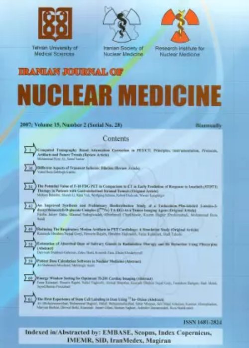
فهرست مطالب
Iranian Journal of Nuclear Medicine
Volume:13 Issue: 1, 2006
- تاریخ انتشار: 1384/10/11
- تعداد عناوین: 8
-
-
[Radiopharmaceutical regulation world wide - the resemblances and the differences]Pages 1-13A Radiopharmaceutical is a radioactive compound used for the diagnosis and treatment of human diseases. Radiopharmaceuticals are among the most highly regulated materials administered to patients because they are controlled both as drugs and as radioactive substances. The use of radiopharmaceuticals for any purpose is governed by regulatory agencies in different countries all over the world. Application of radiopharmaceuticals in humans was almost unregulated until the late 1950s. Since then a progression of regulations have been imposed on the use of these compounds in humans. The United states, United Kingdom, European community, Australia and New Zealand are among the pioneer countries in legislation of regulations concerning the preparation, distribution and use of radiopharmaceuticals. Several regulations, guidelines and reports have been published by federal or professional agencies involved in nuclear pharmacy and nuclear medicine to improve the quality of radiopharmaceutical services. While there are similarities in the backbone of the regulations and guidelines regarding radiopharmaceutical practice worldwide, the comprehensiveness of these regulations varies considerably from country to country and even among states of a country. In order to establish a set of radiopharmaceutical regulations for Iran, reviewing the previously published regulations and guidelines and access their conformity to domestic situations have valuable position and selected acts and topics based on local exigencies may serve as a guide for legislation of the regulations for importation, production and use of radiopharmaceuticals in Iran. The aim of this review is to summarize some of the important aspect of radiopharmaceutical regulations and principal agencies that have regulatory authority over the practice of radiopharmacy.
-
Accuracy of SPECT bone scintigraphy in diagnosis of meniscal tears]Pages 1-6IntroductionScintigraphy has been considered as a useful tool in the assessment of meniscal tears. Our objective was to assess the accuracy of single photon emission tomography (SPECT), using MRI as the gold standard, in the diagnosis of meniscal tears.Materials And MethodsBetween January 2003 and February 2005, 45 patients were studied with SPECT and MRI.ResultsThe respective sensitivity rate, specificity rate, and positive and negative predictive accuracies of SPECT were 78 %, 88 %, 80 %, and 86 %.ConclusionSPECT is a valuable imaging technique and could be considered as competitive for MRI in diagnosis of meniscal tears. SPECT is specially a useful alternative when MRI is unavailable or unsuitable and it is beneficial when more possible accuracy is desired (when MRI results are either inconclusive or contradict with other clinical data).
-
[99mTc Direct radiolabeling of PR81, a new anti-MUC1 monoclonal antibody for radioimmunoscintigraphy]Pages 7-16IntroductionMonoclonal antibodies labeled mainly with 99mTc are being widely used as imaging agents in nuclear medicine. Recently PR81 was introduced as a new murine anti-MUC1 MAb against human breast carcinoma. This antibody reacts with the tandem repeat of 20-mer peptide of protein backbone, and is of IgG1 class, with an affinity of 2.19×10⁻⁸ M-1. Due to high specific reactivity, this antibody was shown to have high diagnostic potential in nuclear medicine. As the first step towards the use of an antibody for imaging purposes it has to be labeled with a radioisotope. In this study we have investigated a method for optimum labeling of this MAb with 99mTc.Materials And MethodsThe Ab reduction was performed with 2-mercaptoethanol (2-ME) at a molar ratio of 2000:1 (2-ME:MAb) and reduced Ab was labeled with 99mTc via stannous tartrate as a transchelator. The labeling efficiency was determined by ITLC. The integrity of reduced MAb was checked by means of SDS-PAGE and gel filtration chromatography (FPLC) on Superose 12 HR 10/30 (purity>99%). The amount of radiocolloids were measured by cellulose nitrate electrophoresis. In vitro stability of labeled product was checked in time intervals over 24 hrs by ITLC. Radioimmunoassay was used to test immunoreactivity of the labeled MAb. Biodistribution of the radiolabeled MAb was studied in normal mice at 4 and 24 hrs post-injection.ResultsThe labeling efficiency was %96±2.8 with high invitro stability (%82±2.7 in 24 hrs) and less than %2 radiocolloids were found. There was no Ab fragmentation due to reduction procedure. Both labeled and unlabeled MAbs were able to compete for binding to the MUC1. Biodistribution studies in normal mice showed that there was no significant accumulation in any organ.ConclusionBecause of no significant loss of immunoreactivity of MAb due to labeling procedure, high in vitro stability and no accumulation in vital organs, 99m-Tc-PR81 may be a promising candidate for radioimmunoscintigraphy studies of breast cancer.
-
Pages 14-18IntroductionUrinary obstructive disease is one of the most common disorders in urinary system. Dilatation which is detected by sonography, is a key finding in obstruction in most of the cases, also could be found following surgery, lithotripsy and vesico–urethral reflux. Radioistope diuretic washout renography is a useful method to differentiate mechanical and non mechanical obstruction.Materials and Methods20 patients were studied with diuretic renography. They were hydrated and emptied their bladders before study. 99mTc–DTPA was injected in supine position for each patient. Images were obtained in 1 min time interval. Then 40 mg furosmide was injected at minute 16th intravenously and sequential imaging were continued for another 20 minutes. Subsequently time activity curves and images were analyzed.ResultsAmong 20 patients, 8 had urinary stones, 9 uretro-pelvic junction stenosis, 2 posterior urethral valve and one case with single kidney. Results showed 13 mechanical and 7 non – mechanical obstruction. Sensitivity of the procedure was 85%.ConclusionThis study revealed that diuretic radionuclide renography is a sensitive and simple method for diagnosis of urinary obstruction.
-
Pages 19-27In this study, the preparation of 111In-Bleomycin complex was optimized for temperature, time, concentration and pH during several experiments. The results showed that by the addition of bleomycin to 111In-InCl3 sample (dried under flow of N2) and heating the mixture up to 90-100°C the radiopharmaceutical can be obtained in 99.5% yield and a specific activity of 1.745 Ci/mmol. The final sample was injected into fibrosarcoma-bearing mice via their tail vein. Satisfactory preliminary SPECT images were acquired between 2-4 hours post-injection showing suitable accumulation in fibrosarcoma tumors.
-
Pages 27-32IntroductionThis study evaluated the diagnostic value of the dipyridamole induced electrocardiuogram (ECG) cahnges, comparing with myocardial perfusion imaging.Materials And Methods222 patinets were studied with dipyridamole infusion (based on ECG criteria) as well as Dipyridamole-myocardial perfusion imaging (MPI).ResultsAbnormal dipyridamole test and abnormal MPI were noted in 11% and 56.3% of patients, respectively. Of 25 postive dip. tests, 20 patients (80%) showed abnormal MPI, more commonly (43.3%) as fixed lesion associated with ischemia. Of dipyridamole negative group, 53.3% showed abnormal MPI. Normal MPI was consistent with normal dipyridamole test in about 94%, while in presence of abnormal MPI, 16% had abnormal dipyridamole test.ConclusionPositive dipyridamole test more likely implies abnormal MPI. More severe CAD in more frequency of positive dipyridamole test. Coronary artery disease (CAD) could not be reliably excluded in presence of normal dipyridamole stress test.
-
Pages 32-37IntroductionMotion of the patient during myocardial perfusion SPECT could potentially results in false perfusion defects. The effect of different reconstruction methods on these artifacts is not studied. Clarification of the relation between the extent, severity and duration of motion with the resultant artifacts may be helpful in designing special soft wares for motion correction. This study is a preliminary evaluation of motion artifacts in different reconstruction methods.Materials And MethodsNormal myocardial perfusion SPECT from a patient with low risk of CAD and no motion during imaging according to the review of cinematic images by 3 nuclear medicine specialists is selected. The scan was acquired one hour after IV injection of 740MBq of Tc-99m-MIBI through 180 degrees from RAO 45 to LPO 45, in 32 frames and 30 seconds per frame. The original images were moved artificially in frames 7, 16 and 24 (as initial, mid and late portion of imaging) from 1 to 3 pixels and for 30-90 seconds using VISION software. Also this artificial motion was applied in X and Y axis and in negative and positive directions. One hundred forty four images were produced and all were reconstructed using filtered back projection (FBP) and iterative (OSEM) reconstructions and interpreted visually and semi-quantitatively (using 17 segment model with 5 point scoring).ResultsApplying one, two and three pixels motion converted normal scan to abnormal image in 2.8%, 25% and 39.6% respectively using FBP and in 18.8%, 43.2% and 68.8% respectively using OSEM reconstruction.(P<0.001). Correlation of interpretation (Kappa) for two sets of images (FBP vs OSEM) was 0.488. Summed score (SS) for each scan reflects the intensity and severity of the defects. Overall, mean summed score was 5.25 in FBP and 8.08 in OSEM reconstruction (P<0.05). Mean SS in returning motions for 1, 2 and 3 frames(corresponding to 30, 60 and 90 seconds) were 2.52, 6 and 8.1 using FBP respectively and 3.85, 8.77 and 11.58 in OSEM respectively (ANOVA, P<0.001). With non-returning motions no normal image was noted in both reconstruction methods.ConclusionThis study showed that OSEM reconstruction is more sensitive for motion artifacts compared to FBP. Also severity and extent of the defects were larger in OSEM reconstruction compared to FBP. Severity of the artifacts is increased with increasing duration of motion (or number of frames involved).
-
Pages 38-51In this study, 16O(p,α)13N nuclear reaction was chosen as the best method of N-13 production. High purity natural water targets were bombarded by 18 MeV protons with an intensity of 8μA. A production yield of 13.5 mCi/μAh was obtained. Chemical modification of 13N-nitrogen oxides into [13N]NH3 was accomplished using De Varda''s catalyst. Chemical, radiochemical and radionuclide purity controls performed on the final product showed high radiopharmaceutical purity (>98%) in the form of [13N]NH3. [13N]NH3 was formulated in normal saline media at appropriate pH and sterile conditions. Microbial and LAL tests were performed on the final product in order to assure its safety for administration to experimental subjects.


