فهرست مطالب

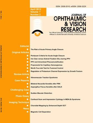
Journal of Ophthalmic and Vision Research
Volume:7 Issue: 2, Apr-Jun 2012
- تاریخ انتشار: 1391/05/08
- تعداد عناوین: 14
-
-
Page 111PurposeTo compare anterior segment and ocular biometric parameters in unaffected fellow eyes of patients with a previous attack of acute angle closure (AAC), primary angle closure suspect (PACS) eyes, and normal eyes; and to identify eyes at high risk of AAC among primary angle closure suspects.MethodsIn this case-control study, 16 unaffected fellow eyes of patients with a previous attack of AAC (group I), 20 PACS eyes (group II) and 18 normal eyes (group III) underwent Pentaca and A-scan echography.ResultsMean anterior chamber volume was 72±18, 77±18 and 176±44? l in groups I, II, and III, respectively (P)
-
Page 118PurposeTo compare the outcomes of iris claw anterior chamber intraocular lens (IC-ACIOL) with that of scleral fixation posterior chamber intraocular lens (SF-PCIOL) implantation during pars plana vitrectomy (PPV) as initial surgery to correct aphakia.MethodsTwelve patients with complicated cataract surgery or trauma who had suffered nucleus, whole crystalline lens or intraocular lens (IOL) drop into the vitreous cavity, and undergone PPV with IC-ACIOL implantation over a period of one year were evaluated for the purpose of this study. Uncorrected visual acuity (UCVA), best corrected visual acuity (BCVA), central corneal thickness (CCT), spherical equivalent (SE) refractive error, astigmatism and complications were recorded. The results were compared to outcomes of another group of 13 patients who had previously undergone PPV with SF-PCIOL implantation.ResultsMean improvement of UCVA was greater in IC-ACIOL eyes as compared to the SF-PCIOL group (-1.17±0.28 versus -0.89±0.21 logMAR, P=0.01), corresponding values for postoperative BCVA were 0.24±0.17 and 0.44±0.22 logMAR (P=0.041), respectively. Average postoperative SE was comparable in the IC-ACIOL and SF-PCIOL groups at 0.6±1.03 and 0.56±1.23 diopters, respectively (P=0.290). However, 10 (83.3%) IC-ACIOL eyes versus 6 (46.1%) SF-PCIOL eyes had SE within 1 diopter of emmetropia (P=0.048). Mean postoperative increase in CCT was comaparble between the study groups (P=0.126).ConclusionIn the absence of sufficient capsular support, the use of an IC-ACIOL for correction of aphakia during PPV can be a good alternative and seems to entail better visual outcomes as compared to SF-PCIOL.
-
Page 125PurposeTo report the outcomes of pars plana vitrectomy (PPV) and intravitreal phacoemulsification in patients with dropped nuclei/nuclear fragments following complicated cataract surgery.MethodsIn this retrospective case series, charts of patients who had undergone PPV and intravitreal phacoemulsification for removal of dislocated nuclei/lens fragments were reviewed. After standard PPV, a conventional phacoemulsification probe with an amputated sleeve was used for grasping and emulsifying the nucleus/nuclear fragments in mid/anterior vitreous cavity. Pre- and postoperative visual acuity, and intra- and postoperative complications were recorded.ResultsA total of 22 patients with mean age of 71.1±8.2 years were studied. Mean interval between complicated cataract surgery and PPV was 26.6±36.5 (range: 0-120) days. Patients were followed for a mean of 105.5±57.5 days. Preoperatively, best corrected visual acuity was 2.4±0.6 logMAR which was improved to 1.4±0.6 logMAR at final follow-up (P
-
Page 130PurposeTo report the long-term results of treatment of pediatric capillary hemangiomas with oral propranolol.MethodsThree infants, 3 to 4 months of age, with periocular capillary hemangiomas were treated with oral propranolol solution (Inderal, 20mg/5ml) 2-3 mg/kg per day divided in 2 doses. Propranolol was continued up to the end of the first year of life and tapered over 2-3 weeks. All infants were followed for 20 months. Lesion size and evolution were assessed during the follow-up period.ResultsSignificant improvement was noted in all patients in the first 2 months of therapy with slow and continuous effect throughout the follow-up period. No serious complications were observed.ConclusionOral propranolol can be used as a first line agent in children with capillary hemangiomas.
-
Page 134PurposeTo compare the results of Worth 4-dot test (WFDT) performed in dark and light, and at different distances, with fusional control in patients with intermittent exotropia (IXT).MethodsDark and light WFDT was performed for new IXT subjects at different distances and the results were compared with level of office-based fusional control.ResultsFifty IXT patients including 17 male and 33 female subjects participated in the study. A significant difference (P)
-
Page 139PurposeTo evaluate the role of brain derived neurotrophic factor (BDNF) and superior collicular extract (SCE) on the expression of the 1.6 subfamily of voltage-gated potassium channels (VG Kv 1.6 channels) in retinal ganglion cells (RGCs) of rats in an in vitro model.MethodsNeonatal retinal cultures were supplemented with trophic factors of interest, namely BDNF and SCE, at 0 DIV (days in vitro), 6 DIV and both 0 and 6 DIV. The expression of VG Kv 1.6 channels was evaluated by immunostaining with anti Kv 1.6 and immunofluorescence was measured by confocal scanning laser microscopy on 4, 6, 8, 10 and 12 DIV. The immunofluorescence indirectly measured the quantity of ion channels being expressed.ResultsRGCs were identified by their soma size. BDNF and SCE enhanced RGC survival by enhancing extensive neurite outgrowth, and increased the expression of VG Kv 1.6 channels; the effect of SCE was more significant than BDNF. Trophic factors also enhanced the survival of RGCs by increasing the expression of ion channels thereby contributing to spontaneous bursts of action potentials in the early stages of RGC development.ConclusionThe expression of delayed rectifier VG Kv 1.6 channels in RGCs may determine membrane excitability and responsiveness to trophic factors, this plays a key role in the refinement of developing retinal circuits.
-
Page 148The advent of new technologies such as high definition optical coherence tomography (OCT) has not only provided unprecedented imaging capabilities, but also raised the need to define concepts not yet settled and often confusing such as the vitreomacular traction (VMT) syndrome. While technological advances drive us into the future by clarifying the pathophysiology of many diseases and enabling novel therapeutic options, it is at the same time necessary to review basic disease concepts in addition to definitions and classifications. VMT syndrome is implicated in the pathophysiology of a number of macular disorders, translating into a variety of anatomical and functional consequences underscoring the complexity of the condition. These macular changes are closely related to the VMT configuration and have led to proposing classification of this syndrome based on OCT findings. The size and severity of the remaining vitreomacular attachment may define the specific maculopathy. Focal VMT usually leads to macular hole formation, tractional cystoid macular edema and foveal retinal detachment, while broad VMT is associated with epiretinal membranes, diffuse retinal thickening and impaired foveal depression recovery. Despite similar postoperative visual acuity (VA) in focal and broad VMT subgroups, visual improvement is greater with focal VMT because preoperative VA is frequently lower. Surgical procedures are effective to relieve VMT and improve VA in most eyes; outcomes vary with VMT morphology and the duration of symptoms.
-
Page 162PurposeTo report the clinical, confocal scan, and histopathologic features of nocardia keratitis in a patient who developed bilateral infection following photorefractive keratectomy (PRK). Case Report: A 23-year-old woman underwent bilateral PRK for low myopia. On postoperative day 3, dense central stromal infiltrates were noticed in both eyes. Empirical antibiotic therapy was initiated which was converted into specific therapy after a definite diagnosis was made based on clinical features and confirmed by confocal scan and histopathologic findings. Clinical and confocal scan features were consistent with the diagnosis of Nocardia keratitis, and topical 2% amikacin eye drops were started. Because of poor response to medical therapy, lamellar keratectomy was performed in both eyes which shortened the treatment course. Histopathologic examination reconfirmed the initial diagnosis.ConclusionFamiliarity with clinical and confocal scan features facilitates early diagnosis of Nocardia keratitis leading to proper management and hence a rapid therapeutic response.
-
Page 180PurposeTo report the clinical, confocal scan, and histopathologic features of nocardia keratitis in a patient who developed bilateral infection following photorefractive keratectomy (PRK). Case Report: A 23-year-old woman underwent bilateral PRK for low myopia. On postoperative day 3, dense central stromal infiltrates were noticed in both eyes. Empirical antibiotic therapy was initiated which was converted into specific therapy after a definite diagnosis was made based on clinical features and confirmed by confocal scan and histopathologic findings. Clinical and confocal scan features were consistent with the diagnosis of Nocardia keratitis, and topical 2% amikacin eye drops were started. Because of poor response to medical therapy, lamellar keratectomy was performed in both eyes which shortened the treatment course. Histopathologic examination reconfirmed the initial diagnosis.ConclusionFamiliarity with clinical and confocal scan features facilitates early diagnosis of Nocardia keratitis leading to proper management and hence a rapid therapeutic response.

