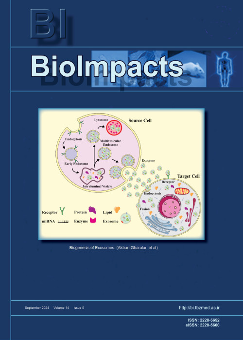
فهرست مطالب
Biolmpacts
Volume:13 Issue: 1, Jan 2023
- تاریخ انتشار: 1401/10/22
- تعداد عناوین: 8
-
-
Pages 1-3
The delivery of chemotherapies to brain tumors faces the difficult task of crossing the blood-brain barrier (BBB).1-4 The brain capillary endothelial cells (BCECs) along with other cell lines, such as astrocytes and pericytes, form the BBB. This highly selective semipermeable barrier separates the blood from the brain parenchyma. The BBB controls the movement of drug molecules in a selective manner5 and maintains central nervous system (CNS) homeostasis. Depending on the properties of drugs such as their hydrophilic-lipophilic balance (HLB), some can cross the BBB through passive diffusion.6 However, this approach alone has not led to successful drug developments due to low net diffusion rates and systemic toxicity. Although the use of nanomedicine has been proposed to overcome these drawbacks, many recent studies still rely on the so-called ‘enhanced permeability and retention (EPR)’ effect though there is a realization in the field of drug delivery that EPR effect may not be sufficient for successful drug delivery to brain tumors. Since, compared to many other solid tumors, brain tumors pose additional challenges such as more restrictive blood-tumor barrier as well as the well-developed lymphatic drainage, the selection of functional moieties on the nanocarriers under consideration must be carried out with care to propose better solutions to this challenge.
Keywords: Blood-brain-barrier, Targeted delivery, EPR effect, Lymphatic drainage, Brain tumors, Nanomedicine -
Pages 5-16Introduction
Here, the interaction behavior between propyl acridones (PA) and calf thymus DNA (ct-DNA) has been investigated to attain the features of the binding behavior of PA with ct-DNA, which includes specific binding sites, modes, and constants. Furthermore, the effects of PA on the conformation of ct-DNA seem to be quite significant for comprehending the medicine’s mechanism of action and pharmacokinetics.
MethodsThe project was accomplished through means of absorbance studies, fluorescence spectroscopy, circular dichroism, viscosity measurement, thermal melting, and molecular modeling techniques.
ResultsThe intercalation of PA has been suggested by fluorescence quenching and viscosity measurements results while the thermal melting and circular dichroism studies have confirmed the thermal stabilization and conformational changes that seem to be associated with the binding. The binding constants of ct-DNA-PA complex, in the absence and presence of EMF, have been evaluated to be 6.19 × 10⁴ M-1 and 2.95 × 10⁴ M-1 at 298 K, respectively. In the absence of EMF, the ∆H0 and ∆S0 values that occur in the interaction process of PA with ct-DNA have been measured to be -11.81 kJ.mol-1 and 51.01 J.mol-1K-1, while in the presence of EMF they were observed to be -23.34 kJ.mol-1 and 7.49 J.mol-1K-1, respectively. These numbers indicate the involvement of multiple non-covalent interactions in the binding procedure. In a parallel study, DNA-PA interactions have been monitored by molecular dynamics simulations; their results have demonstrated DNA stability with increasing concentrations of PA, as well as calculated bindings of theoretical ΔG0.
ConclusionThe complex formation between PA and ct-DNA has been investigated in the presence and absence of EMF through the multi spectroscopic techniques and MD simulation. These findings have been observed to be parallel to the results of KI and NaCl quenching studies, as well as the competitive displacement with EB and AO. According to thermodynamic parameters, electrostatic interactions stand as the main energy that binds PA to ct-DNA. Regarding the cases that involve the Tm of ct-DNA, EMF has proved to increase the stability of binding between PA and ct-DNA.
Keywords: ct-DNA, Propyl acridon, Spectroscopy, Intercalator, Molecular dynamics, Anti-cancer drug -
Pages 17-29Introduction
The present study was done to assess the effect of molecularly-targeted core/shell of iron oxide/gold nanoparticles (Fe3O4@AuNPs) on tumor radiosensitization of SKBr-3 breast cancer cells.
MethodsHuman epidermal growth factor receptor-2 (HER-2)-targeted Fe3O4@AuNPs were synthesized by conjugating trastuzumab (TZ, Herceptin) to PEGylated (PEG)-Fe3O4@AuNPs (41.5 nm). First, the Fe3O4@Au core-shell NPs were decorated with PEG-SH to synthesize PEG-Fe3O4@AuNPs. Then, the TZ was reacted to OPSS-PEG-SVA to conjugate with the PEG-Fe3O4@AuNPs. As a result, structure, size and morphology of the developed NPs were assessed using Fourier-transform infrared (FT-IR) spectroscopy, dynamic light scattering (DLS) and transmission electron microscopy (TEM), and ultraviolet-visible spectroscopy. The SKBr-3 cells were treated with different concentrations of TZ, Fe3O4@Au, and TZ-PEG-Fe3O4@AuNPs for irradiation at doses of 2, 4, and 8 Gy (from X-ray energy of 6 and 18 MV). Cytotoxicity was assessed by MTT assay, BrdU assay, and flow cytometry.
ResultsResults showed that the targeted TZ-PEG-Fe3O4@AuNPs significantly improved cell uptake. The cytotoxic effects of all the studied groups were increased in a higher concentration, radiation dose and energy-dependent manner. A combination of TZ, Fe3O4@Au, and TZ-PEG-Fe3O4@AuNPs with radiation reduced cell viability by 1.35 (P=0.021), 1.95 (P=0.024), and 1.15 (P=0.013) in comparison with 8 Gy dose of 18 MV radiation alone, respectively. These amounts were obtained as 1.27, 1.58, and 1.10 for 8 Gy dose of 6 MV irradiation, respectively.
ConclusionRadiosensitization of breast cancer to mega-voltage radiation therapy with TZ-PEG-Fe3O4@AuNPs was successfully obtained through an optimized therapeutic approach for molecular targeting of HER-2.
Keywords: Breast cancer, Radiation therapy, Active targeting, Trastuzumab, Au nanoparticles -
Pages 31-42Introduction
Treatment of critical-sized bone defects is challenging. Tissue engineering as a state-of-the-art method has been concerned with treating these non-self-healing bone defects. Here, we studied the potentials of new three-dimensional nanofibrous scaffolds (3DNS) with and without human adipose mesenchymal stem cells (ADSCs) for reconstructing rat critical-sized calvarial defects (CSCD).
MethodsScaffolds were made from 1- polytetrafluoroethylene (PTFE), and polyvinyl alcohol (PVA) (PTFE/ PVA group), and 2- PTFE, PVA, and graphene oxide (GO) nanoparticle (PTFE/ PVA/GO group) and seeded by ADSCs and incubated in osteogenic media (OM). The expression of key osteogenic proteins including Runt-related transcription factor 2 (Runx2), collagen type Iα (COL Iα), osteocalcin (OCN), and osteonectin (ON) at days 14 and 21 of culture were evaluated by western blot and immunocytochemistry methods. Next, 40 selected rats were assigned to five groups (n=8) to create CSCD which will be filled by scaffolds or cell-containing scaffolds. The groups were denominated as the following order: Control (empty defects), PTFE/PVA (PTFE/PVA scaffolds implant), PTFE/PVA/GO (PTFE/PVA/GO scaffolds implant), PTFE/PVA/Cell group (PTFE/PVA scaffolds containing ADSCs implant), and PTFE/PVA/GO/Cell group (PTFE/PVA/GO scaffolds containing ADSCs implant). Six and 12 weeks after implantation, the animals were sacrificed and bone regeneration was evaluated using computerized tomography (CT), and hematoxylin-eosin (H&E) staining.
ResultsBased on the in-vitro study, expression of bone-related proteins in ADSCs seeded on PTFE/PVA/GO scaffolds were significantly higher than PTFE/PVA scaffolds and TCPS (P<0.05). Based on the in-vivo study, bone regeneration in CSCD were filled with PTFE/PVA/GO scaffolds containing ADSCs were significantly higher than PTFE/PVA scaffolds containing ADSCs (P<0.05). CSCD filled with cell-seeded scaffolds showed higher bone regeneration in comparison with CSCD filled with scaffolds only (P<0.05).
ConclusionThe data provided evidence showing new freeze-dried nanofibrous scaffolds formed from hydrophobic (PTFE) and hydrophilic (PVA) polymers with and without GO provide a suitable environment for ADSCs due to the expression of bone-related proteins. ADSCs and GO in the implanted scaffolds had a distinct effect on the bone regeneration process in this in-vivo study.
Keywords: Calvarial defect, Three dimensional nanofibrous scaffold, Polyvinyl alcohol, Polytetrafluoroethylene, Graphene oxide nanoparticle, Human adipose mesenchymal stem cells -
Pages 43-50Introduction
The current experiment aimed to address the impact of type 2 diabetes mellitus on autophagy status in the rat pulmonary tissue.
MethodsIn this study, 20 male Wistar rats were randomly allocated into two groups as follows: control and diabetic groups. To induce type 2 diabetes mellitus, rats received a combination of streptozotocin (STZ) and a high-fat diet. After confirmation of diabetic condition, rats were maintained for 8 weeks and euthanized for further analyses. The pathological changes were assessed using H&E staining. We also measured transforming growth factor-β (TGF-β), bronchoalveolar lavage fluid (BALF), and tumor necrosis factor-α (TNF-α) in the lungs using ELISA and real-time PCR analyses, respectively. Malondialdehyde (MDA) and superoxide dismutase (SOD) levels were monitored in diabetic lungs to assess oxidative status. We also measured the expression of becline-1, LC3, and P62 to show autophagic response under diabetic conditions. Using immunofluorescence staining, protein levels of LC3 was also monitored.
ResultsH&E staining showed pathological changes in diabetic rats coincided with the increase of TNF-α (~1.4-fold) and TGF-β (~1.3-fold) compared to those in the normal rats (P < 0.05). The levels of MDA (5.6 ± 0.4 versus 6.4 ± 0.27 nM/mg protein) were increased while SOD (4.2 ± 0.28 versus 3.8 ± 0.13 U/mL) activity decreased in the diabetic rats (P < 0.05). Real-time polymerase chain reaction (PCR) analysis showed the up-regulation of Becline-1 (~1.35-fold) and LC3 (~2-fold) and down-regulation of P62 (~0.8-fold) (P < 0.05), showing incomplete autophagic flux. We noted the increase of LC3+ cells in diabetic condition compared to that in the control samples.
ConclusionThe prolonged diabetic condition could inhibit the normal activity of autophagy flux, thereby increasing pathological outcomes.
Keywords: Autophagy, Inflammation, Pulmonary tissue, Type 2 diabetes mellitus -
Pages 51-61Introduction
Silibinin is a natural flavonoid compound known to induce apoptosis in cancer cells. Despite silibinin's safety and efficacy as an anticancer drug, its effects on inducing immunogenic cell death (ICD) are largely unknown. Herein, we have evaluated the stimulating effects of silibinin on ICD in cancer cells treated with silibinin alone or in combination with chemotherapy.
MethodsThe anticancer effect of silibinin, alone or in combination with doxorubicin or oxaliplatin (OXP), was assessed using the MTT assay. Compusyn software was used to analyze the combination therapy data. Western blotting was conducted to examine the level of STAT3 activity. Flow cytometry was used to analyze calreticulin (CRT) and apoptosis. The heat shock protein (HSP70), high mobility group box protein1 (HMGB1), and IL-12 levels were assessed by ELISA.
ResultsCompared to the negative control groups, silibinin induced ICD in CT26 and B16F10 cells and significantly enhanced the induction of this type of cell death by doxorubicin, and these changes were allied with substantial increases in the level of damage-associated molecular patterns (DAMPs) including CRT, HSP70, and HMGB1. Furthermore, conditioned media from cancer cells exposed to silibinin and doxorubicin was found to stimulate IL-12 secretion in dendritic cells (DCs), suggesting the link of this treatment with the induction of Th1 response. Silibinin did not augment the ICD response induced by OXP.
ConclusionOur findings showed that silibinin can induce ICD and it potentiates the induction of this type of cell death induced by chemotherapy in cancer cells.
Keywords: Immunotherapy, Combination therapy, DAMPs, Silibinin, Th1 response -
Pages 63-72Introduction
Biocompatible and biodegradable scaffolds based on natural polymers such as gelatin and chitosan (CS) provide suitable microenvironments in dental tissue engineering. In the present study, we report on the synthesis of injectable thermosensitive hydrogel (PNIPAAm-g-CS copolymer/gelatin hybrid hydrogel) for osteogenic differentiation of human dental pulp stem cells (hDPSCs).
MethodsThe CS-g-PNIPAAm was synthesized using the reaction of carboxyl terminated PNIPAAm with CS, which was then mixed with various amounts of gelatin solution in the presence of genipin as a chemical crosslinker to gain a homogenous solution. The chemical composition and microstructures of the fabricated hydrogels were confirmed by FT-IR and SEM analysis, respectively. To evaluate the mechanical properties (e.g., storage and loss modulus of the gels), the rheological analysis was considered. Calcium deposition and ALP activity of DPSCs were carried out using alizarin red staining and ALP test. While the live/dead assay was performed to study its toxicity, the real-time PCR was conducted to investigate the osteogenic differentiation of hDPSCs cultured on prepared hydrogels.
ResultsThe hydrogels with higher gelatin incorporation showed a slightly looser network compared to the other ones. The hydrogel with less gelatin indicates a rather higher value of G', indicating a higher elasticity due to more crosslinking reaction of amine groups of CS via a covalent bond with genipin. All the hydrogels contained viable cells with negligible dead cells, indicating the high biocompatibility of the prepared hydrogels for hDPSCs. The quantitative results of alizarin red staining displayed a significant rise in calcium deposition in hDPSCs cultured on prepared hydrogels after 21 days. Further, hDPSCs cultured on hydrogel with more gelatin displayed the most ALP activity. The expression of late osteogenic genes such as OCN and BMP-2 were respectively 6 and 4 times higher on the hydrogel with more gelatin than the control group after 21 days.
ConclusionThe prepared PNIPAAm-g-CS copolymer/gelatin hybrid hydrogel presented great features (e.g., porous structure, suitable rheological behavior, and improved cell viability), and resulted in osteogenic differentiation necessary for dental tissue engineering.
Keywords: Injectable hydrogel, Chitosan, Gelatin, Dental stem cells, Regenerative medicine, Tissue engineering -
Pages 73-84Introduction
The mixed flavonoid supplement (MFS) [Trimethoxy Flavones (TMF) + epigallocatechin-3-gallate (EGCG)] can be used to suppress inflammatory ulcers as an ethical medicine in Ayurveda. The inflammation of the rectum and anal regions is mostly attributed to nuclear factor kappa beta (NF-κB) signaling. NF-κB stimulates the expression of matrix metalloproteinase (MMP9), inflammatory cytokines tumor necrosis factor (TNF-α), and interleukin-1β (IL-1β). Although much research targeted the NF-κB and MMP9 signaling pathways, a subsequent investigation of target mediators in the inflammatory ulcer healing and NF-κB pathway has not been done.
MethodsThe docking studies of compounds TMF and EGCG were performed by applying PyRx and available software to understand ligand binding properties with the target proteins. The synergistic ulcer healing and anti-arthritic effects of MFS were elucidated using dextran sulfate sodium (DSS)-induced colon ulcer in Swiss albino rats. The colon mucosal injury was analyzed by colon ulcer index (CUI) and anorectic tissue microscopy. The IL-1β, tumor necrosis factor (TNF-α), and the pERK, MMP9, and NF-κB expressions in the colon tissue were determined by ELISA and Western blotting. RT-PCR determined the mRNA expression for inflammatory marker enzymes.
ResultsThe docking studies revealed that EGCG and TMF had a good binding affinity with MMP9 (i.e., -6.8 and -6.0 Kcal/mol) and NF-kB (-9.4 and 8.3 kcal/mol). The high dose MFS better suppressed ulcerative colitis (UC) and associated arthritis with marked low-density pERK, MMP9, and NF-κB proteins. The CUI score and inflammatory mediator levels were suppressed with endogenous antioxidant levels in MFS treated rats.
ConclusionThe MFS effectively unraveled anorectic tissue inflammation and associated arthritis by suppressing NF-κB-mediated MMP9 and cytokines.
Keywords: MFS, Anorectic ulcer, Arthritis, NF-κB, MMP9, Cytokines


